
Introduction
Restoration of the original anatomy is a crucial aspect of fracture treatment. During surgery, coronal and sagittal angulations in long bones can be aligned using two-dimensional (2D) fluoroscopic imaging. However, assessing rotational deformity with conventional 2D fluoroscopic images is subjective. While the extent of fragment rotation can be estimated by comparing the affected side to the contralateral normal side, this method necessitates images of the contralateral side and relies entirely on its condition. Additionally, bilateral bone morphology is not symmetrical. In cases involving both sides, comparison is not possible, making quantitative estimation of rotation extent unattainable. The intraoperative three-dimensional (3D) estimation of bone position using 2D images has garnered interest in orthopedic surgery, including computer-assisted techniques. Rotation assessment in the lower extremity, such as the femur or tibia, is typically performed using specific landmarks. For instance, the size of the lesser trochanter is used as an indicator of the femur's internal rotation status [1,2]. In contrast, studies on humeral rotation are less common than those for the lower extremity. To date, only a handful of studies have investigated the measurement of humeral rotation without the use of landmarks [3–5]. Therefore, we developed a method to estimate and evaluate the rotational alignment of the proximal humerus using a specific landmark.
The aim of the current study was to quantify the relationship between proximal humeral rotation and the lateral border of the bicipital groove as seen on fluoroscopic imaging. We hypothesized that the lateral border of the bicipital groove could act as a practical landmark for assessing humeral rotation.
Methods
It is not a human population study; therefore, neither approval by the institutional review board nor obtainment of informed consent was required.
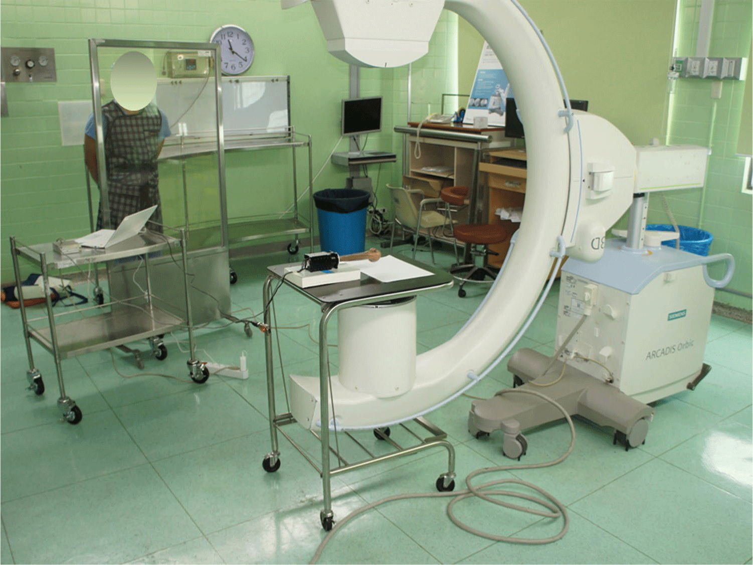
A composite sawbone model of a humerus (#3404, Sawbones, Vashon Island, WA, USA) was sectioned at the midpoint of the shaft. Prior to sectioning, a longitudinal line was drawn on the anterior surface to ensure that the proximal half retained half of this line. The proximal segment of the humeral model was then secured to a custom rotation device, aligning the longitudinal line with the 0° rotation mark on the device. Consequently, a 0° rotation on the device corresponded to a neutral alignment of the proximal segment relative to the distal segment of the humerus. For precise control and high accuracy, we employed a modular actuator with 0.1-mm precision (Dynamixel Pro, ROBOTIS, Seoul, Korea) as the custom rotation device. A metal dot was affixed to the lateral edge of the bicipital groove at the point corresponding to the largest diameter of the humeral head in preparation for fluoroscopic imaging. Since the humeral head is spherical, any point on its surface can serve as a rotational reference through geometric calculation. We chose the lateral edge of the bicipital groove as this reference due to its relative ease of identification on imaging. The location for the metal dot was specifically chosen because the maximum circumference of the hypothetical sphere would exhibit the greatest change with each degree of rotation, thus providing the highest sensitivity to rotational changes. The assembly was positioned on a radiolucent table beneath an image intensifier for imaging purposes. The rotation device was set up to maintain the distal segment stationary while the proximal segment was rotated from −60° to 60° in 5° increments and from −15° to 15° in 1° increments, achieving an accuracy of 0.1°. A fluoroscopic image was captured at each incremental position of rotation (Fig. 1).
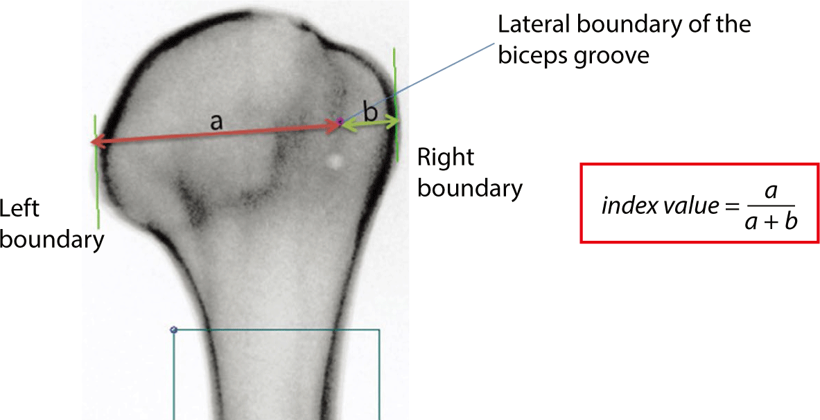
We used a specialized program to calculate an index value indicative of humeral rotation. The user identified a rectangle and three points on fluoroscopic images, as depicted in Fig. 2. The rectangle was placed over the diaphysis to establish the humerus's long axis, which was determined from the selected area through principal component analysis. The medial and lateral boundaries of the humerus were marked, ensuring that both were tangential to its long axis. Additionally, a point was marked at the lateral edge of the bicipital groove. The value “a” was the distance between the medial boundary of the humeral head, “b” was the distance between the lateral boundary of the bicipital groove, and the value of “a+b” was the distance from the medial to the lateral boundary. The index value was the ratio of “a” to “a+b” in equation 1 (Fig. 1), with the assumption that the index value correlates with humeral rotation.
The Kolmogorov–Smirnov test was used to assess the normality of the distribution. The dataset of index values followed a normal distribution; therefore, Pearson's correlation coefficient was employed to examine the relationship between the index value and the angle of humeral rotation. A regression equation was also derived. The threshold for statistical significance was established at P<0.05. Both descriptive and analytical analyses were performed using SPSS version 15.0 (SPSS, Chicago, IL, USA).
Results
The index value showed a strong positive linear correlation with position during internal rotation of the humerus (correlation [IR]=0.998; P<0.001). Similarly, a moderate positive linear correlation was observed with position during external rotation of the humerus (correlation [ER]=0.693; P<0.001). Notably, within the range of 15° internal rotation to 15° external rotation, the correlation remained strongly positive (correlation [IR15–ER15]=0.991; P<0.001; Fig. 3).
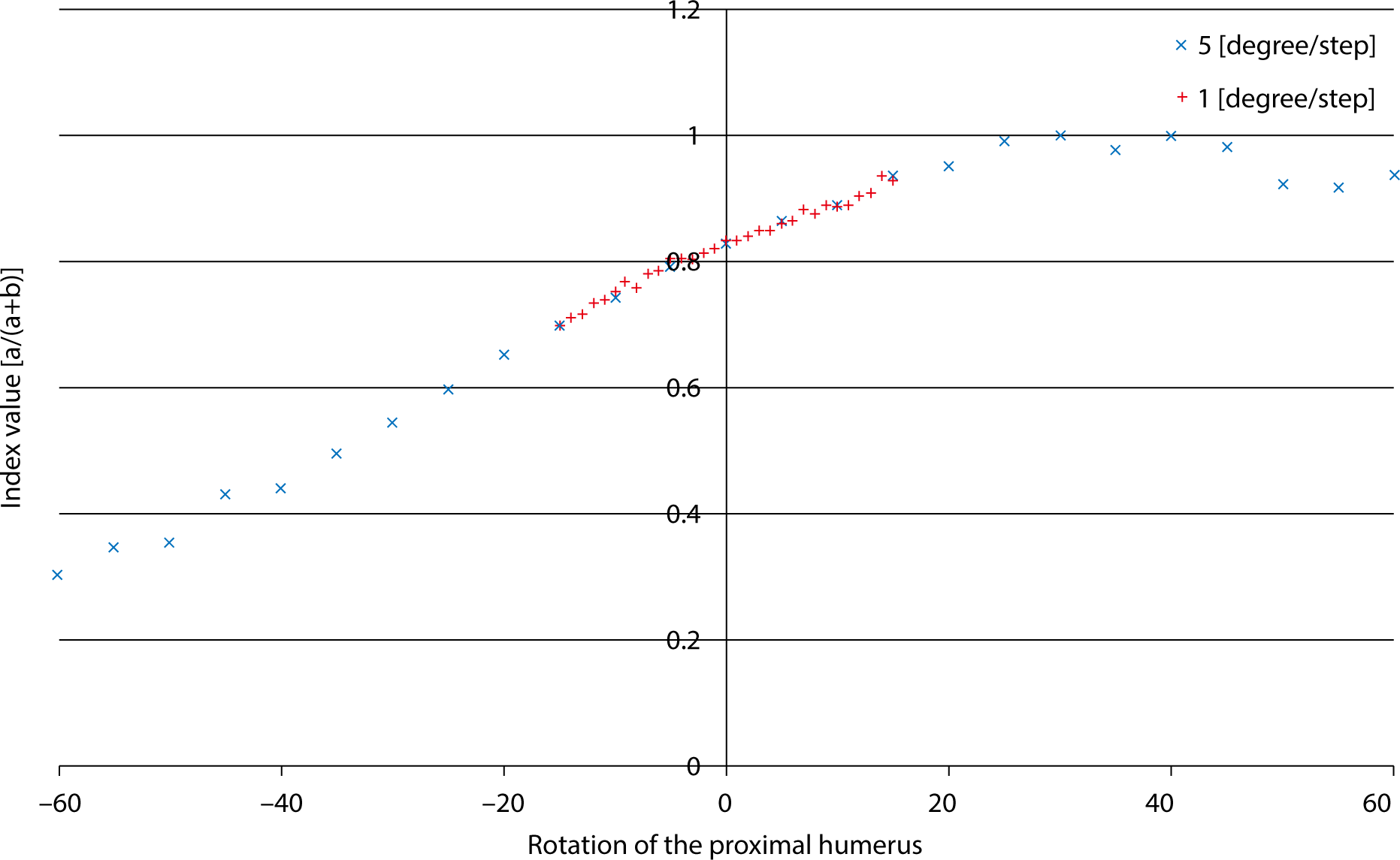
The regression equations for internal and external rotation were as follows:
The regression equation for internal and external rotation between −15° and 15° was as follows:
Index value = 0.00727 × (angle) + 0.82225
Discussion
We found that the index value of the lateral border of the bicipital groove exhibits a moderate-to-strong correlation with the rotational angle of the humerus as seen on fluoroscopic imaging. These findings could prove beneficial for minimally invasive plate osteosynthesis (MIPO), a technique that has recently become more popular.
MIPO offers the advantage of preserving periosteal blood supply; however, it is often associated with rotational malalignment due to the lack of direct visualization of the fracture site [6]. Accordingly, we established a linear correlation between the index landmark and the rotation angle. The clinical significance of our study is that it provides a method for estimating the rotation angle of the proximal humerus. When the distal humerus is positioned neutrally on fluoroscopy, variations in the lateral border of the bicipital groove can indicate the degree of rotation relative to the distal part. This allows for the assessment of rotation without the need for repeated fluoroscopic examination of the distal part.
The acceptable limit for rotational malalignment is generally considered to be 20 degrees. The degree of malrotation is directly related to a reduction in the range of motion [7]. While anatomical rotational alignment is possible using open reduction and internal fixation, achieving correct humeral alignment during MIPO surgery can be more challenging. In acute cases, palpating the epicondyles may be difficult due to traumatic edema or in patients with obesity. Utilizing an index value for measurement provides a quantitative assessment of the reduction and alignment of the fractured fragments. Variations in humeral anatomy among different patients necessitate this approach. By measuring the index value, surgeons can customize the treatment to accommodate individual differences in bone structure and alignment. This is critical for attaining optimal anatomical alignment, which is a key factor in ensuring functional recovery.
Malrotation in the humerus is generally considered more acceptable than in the lower extremities, which has led to limited research on humeral rotation. Consequently, precise criteria or landmarks for assessment using plain radiographs have yet to be established. However, studies by Itoi et al. [8] and Sabo et al. [9] have reported that humeral malrotation leads to malunion, whereas Li et al. [4] found that it had a negative effect on shoulder function. Moreover, recent advances in shoulder and elbow arthroplasty have demonstrated that the sequelae of humeral malrotation are caused by altered kinematics [8,9]. A study on humeral shaft fracture repair assessed rotation during surgery using the cortical step sign [10]. Boileau et al. [11] used the shape of the bicipital groove to assess rotation by comparing the ipsilateral and contralateral sides. However, neither method was able to provide quantitative measurements. CT is highly reliable and accurate for evaluating humeral rotation, but its feasibility during surgery is questionable. Tan et al. [3] used the cortical density of the lesser tuberosity as a landmark for humeral rotation and showed its validity in a cadaver study. In contrast, our study was able to measure rotation in 1° increments using a custom device, thus offering superior accuracy. We utilized the lateral border of the bicipital groove as a landmark because it provides a clear reference point when the lesser tuberosity is not visible, particularly during ranges of internal rotation. A linear correlation was found between the position of this landmark and the degree of humeral rotation. This relationship may need adjustment if the proximal humerus obscures the medial line of the greater tuberosity due to the position of the lesser tuberosity. Nevertheless, within the clinically relevant range of humeral rotation for computer-assisted fracture surgery (internal rotation 15° to external rotation 15°), we observed a strong positive linear correlation with humeral rotation. The accurate estimation of humeral rotation using a landmark is crucial for both conventional MIPO and fracture surgery. The bicipital groove has also been suggested as a reliable intraoperative landmark for restoring humeral retrotorsion during shoulder replacement or for reconstructing the premorbid anatomy of the proximal humerus [12]. Our study confirmed the reliability of using the bicipital groove and found a linear correlation between the landmark and the humeral rotation.
Future trends in orthopedic surgery will rely on robot-assisted or computer-assisted techniques, which can reduce soft tissue damage and increase the accuracy of reduction by targeting the exact point for incision and manipulation [13]. In robot-assisted surgery, exact data points are needed, such as for computer-assisted arthroscopic subscapularis repair. The inability to visualize the subscapularis tendon footprint on arthroscopy is generally accepted. However, careful registration of the palpable lateral border of the bicipital groove allows the surface registration of an anatomical landmark of the proximal humerus. This improves accuracy when inserting an anchor to the lesser tuberosity. We aimed to define accurate landmarks rather than intuitively relying on comparison with the contralateral side. Because the anatomy of the humerus varies and a fragmented or distorted humerus anatomy may hinder the use of this landmark, other complementary 3D methods should be performed to determine the exact position of the proximal humerus. The lateral border of the bicipital groove can be used as a clinically important guide to evaluate humeral rotational alignment for fracture reduction or other computer-assisted surgical procedures, particularly in the range between −15° and 15°, where measurement errors often occur.
Our estimation method has certain limitations. First, the lateral border of the bicipital groove may be obliterated in a comminuted fracture, severe osteoporosis, or the presence of implant-related materials. Improperly positioned shoulder images can also interfere with accurate imaging of the landmark. Thus, the placement of the metal dot may not align with what is observed in fluoroscopic images. However, the lateral border of the bicipital groove becomes more discernible when the humerus is internally rotated. The lesser tubercle can also serve as an intraoperative landmark for humeral rotation. Second, we cannot generalize the data to all patients because of variations in anatomy, such as in the degree of humeral anteversion or anatomical variation of bicipital groove. However, our study demonstrates the value of objective data for estimating humeral rotation, which could be used in a practical clinical setting. Therefore, future studies are needed to determine more generalized or normative data. Finally, estimating the rotation angle using a shoulder image alone assumes that the elbow joint is in its neutral position, which is relatively easy to achieve because of the wide posterior surface. Thus, we assumed that the effect of elbow positioning is minimal.
Conclusion
The lateral border of the bicipital groove can serve as an intraoperative landmark for the quantitative estimation of proximal humeral rotation. This landmark proves beneficial in minimally invasive or robotic surgeries targeting the proximal humerus. Assessing humeral rotation during surgery can enhance the results of humeral fracture repairs and upper arm arthroplasty procedures.