A 44-year-old woman was referred to the cardiology department due to experiencing heaviness in both legs during exertion and decreased ankle-brachial indexes of 0.83 and 0.78, respectively. She had a mild case of aplastic anemia. Lower extremity computed tomographic angiography (CTA) revealed a total occlusion of the left iliac artery from the bifurcation of the abdominal aorta, which was reconstructed at the level of the common femoral artery (
Fig. 1A, red arrow). Atherosclerotic plaque was clearly visible at the abdominal aorta (
Fig. 1B,
C). However, the occluded iliac artery showed thread-like hypoplasia without any definitive atherosclerotic occlusion (
Fig. 1D, red arrow).
Fig. 1. CT angiography findings of the lower extremities. (A) The left iliac artery is completely occluded from the aortic bifurcation (circle; “string of beads” appearance of the right external iliac artery from invasive angiography). (B) Atherosclerotic plaques are present in the abdominal aorta and (C) at the bifurcation site. (D) A thread-like hypoplastic left iliac artery is visible. (E−G) Multiple layers of thread-like stenosis (red arrows) and a beaded appearance of the left popliteal artery are evident on CT angiography (orange arrows).
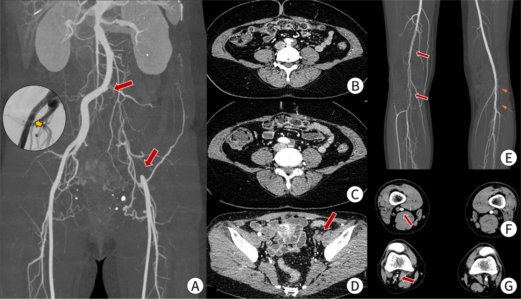
Initially, we suspected congenital atresia of the iliac artery due to the patient's hypoplastic iliac artery, despite this being a rare condition. However, the renal arteries shown on CTA (
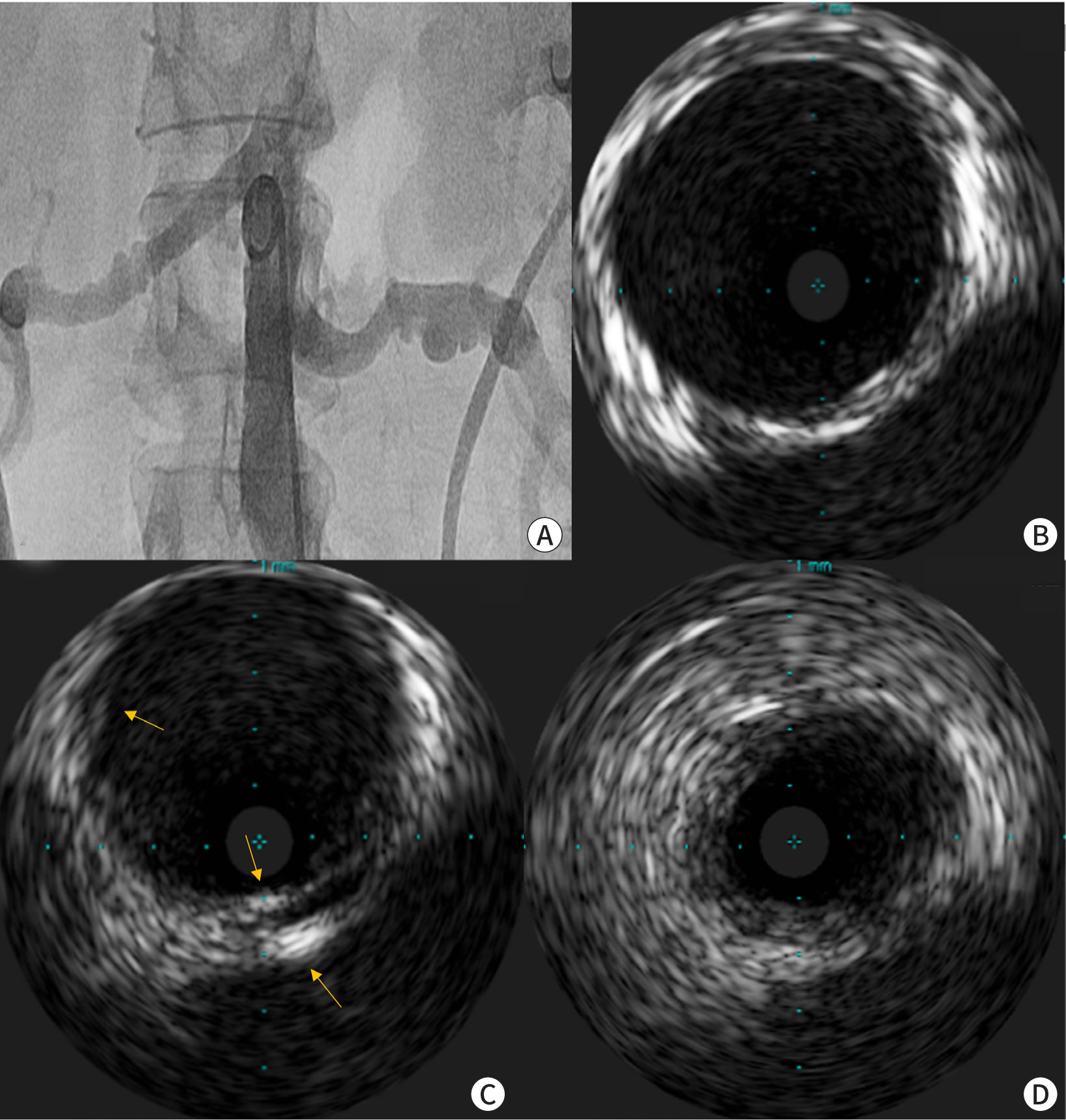
Fig. 1A) suggested the possibility of fibromuscular dysplasia (FMD). We performed angiography on the candidate vessels. Both renal arteries displayed the typical “string of beads” appearance associated with FMD (
Fig. 2A). The findings from intravascular ultrasonography were also consistent with FMD (
Fig. 2B,
C). Both carotid arteries were relatively normal in shape, but had tortuous courses. In contrast to CTA, the right external iliac artery exhibited a “string of beads” appearance on angiography (
Fig. 1A, circle). Additionally, the right popliteal artery showed multiple layers of tread-like stenosis, and the left popliteal artery displayed a beaded appearance on CTA (
Fig. 1E–
G, red arrows).
Fig. 2. Invasive images of the renal arteries. (A) Angiography reveals the typical “string of beads” appearance in both renal arteries. Intravascular ultrasonography indicates varied disease progression. (B) The vessel layers are relatively preserved. (C) There is an intimal fibromuscular ridge and increased medial echogenicity, as indicated by the arrows. (D) Circumferential hyperplasia of the intimal or medial layers is noted, accompanied by denudation of the vessel layers.

FMD is a non-inflammatory and non-atherosclerotic arterial disease of unknown etiology. Historically, FMD was categorized based on the arterial layers affected. However, obtaining pathological specimens is now a rare occurrence. Today, FMD is typically diagnosed through imaging studies, and an angiographic classification is commonly employed [
1].
This patient exhibited tubular and multifocal stenosis, which is characteristic of FMD, and primarily showed lower extremity involvement. This article underscores the importance of considering FMD in middle-aged women presenting with peripheral artery obstructive disease but lacking traditional risk factors. It also highlights the utility of various imaging modalities in such cases.
Acknowledgements
Not applicable.
Conflict of Interest
-
No potential conflict of interest relevant to this article was reported.
Author Contribution
-
The article is prepared by a single author.
Ethics Approval and Consent to Participate
-
Not applicable.
References
- 1. Olin JW, Gornik HL, Bacharach JM, Biller J, Fine LJ, Gray BH, et al. Fibromuscular dysplasia: state of the science and critical unanswered questions: a scientific statement from the American Heart Association. Circulation 2014;129((9)):1048-1078.
Citations
Citations to this article as recorded by