• Contact us
  • E-Submission
ABOUT
BROWSE ARTICLES
JOURNAL POLICIES
FOR CONTRIBUTORS

Articles

Page Path

Images and Solution

Jejunal Mass Presented by Anemia

The Ewha Medical Journal 2011;34(2):64-66. Published online: September 30, 2011

Department of Surgery, Ewha Womans University School of Medicine, Seoul, Korea.

1Department of Pathology, Ewha Womans University School of Medicine, Seoul, Korea.

Corresponding author (ralee@ewha.ac.kr)

Copyright © 2011. Ewha Womans University School of Medicine

This is an Open Access article distributed under the terms of the Creative Commons Attribution Non-Commercial License (http://creativecommons.org/licenses/by-nc/3.0/) which permits unrestricted non-commercial use, distribution, and reproduction in any medium, provided the original work is properly cited.

  • 36 Views
  • 0 Download
prev next
  • 1. Miettinen M, Lasota J. Gastrointestinal stromal tumors--definition, clinical, histological, immunohistochemical, and molecular genetic features and differential diagnosis. Virchows Arch 2001;438:1-12.
  • 2. Yarden Y, Kuang WJ, Yang-Feng T, Coussens L, Munemitsu S, Dull TJ, et al. Human proto-oncogene c-kit: a new cell surface receptor tyrosine kinase for an unidentified ligand. EMBO J 1987;6:3341-3351.
  • 3. Ranchod M, Kempson RL. Smooth muscle tumors of the gastrointestinal tract and retroperitoneum. Cancer 1977;39:255-262.
  • 4. Shiu MH, Farr GH, Papachristou DN, Hajdu SI. Myosarcomas of the stomach: natural history, prognostic factors and management. Cancer 1982;49:177-187.
  • 5. Ueyama T, Guo KJ, Hashimoto H, Daimaru Y, Enjoji M. A clinicopathologic and immunohistochemical study of gastrointestinal stromal tumors. Cancer 1992;69:947-955.
  • 6. Miettinen M, El-Rifai W, Sobin LH, Lasota J. Evaluation of malignancy and prognosis of gastrointestinal stromal tumors: a review. Hum Pathol 2002;33:478-483.
  • 7. Buchdunger E, Cioffi CL, Law N, Stover D, Ohno-Jones S, Bruker BJ, et al. Abl protein-tyrosine kinase inhibitor STI571 inhibits in vitro signal transduction mediated by c-kit and platelet-derived growth factor receptors. J Pharmacol Exp Ther 2000;295:139-145.
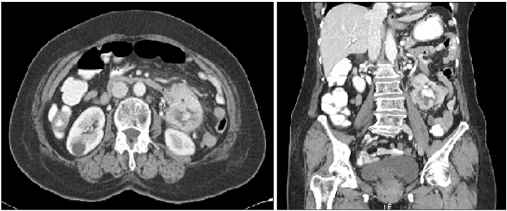
Fig. 1
Abdominal computed tomography findings. 6.7×6.2 cm sized mass with cavitary lesion is visible in the left upper quadrant.
emj-34-64-g001.jpg
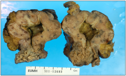
Fig. 2
Macroscopic finding. 8×6 cm sized mass involved the wall of jejunum and the mucosa showed a deep ulceration.
emj-34-64-g002.jpg
Fig. 3
Microscopic findings. (A) Haematoxylin & eosin stain (×400) shows more than 5 atypical mitoses with spindle cell arrangement. (B) Immunohistochemical stain for c-kit (×400) shows nuclear and cytoplasmic positivity in interstitial cells.
emj-34-64-g003.jpg

Figure & Data

References

    Citations

    Citations to this article as recorded by  

      Download Citation

      Download a citation file in RIS format that can be imported by all major citation management software, including EndNote, ProCite, RefWorks, and Reference Manager.

      Format:

      Include:

      Jejunal Mass Presented by Anemia
      Ewha Med J. 2011;34(2):64-66.   Published online September 30, 2011
      Download Citation
      Download a citation file in RIS format that can be imported by all major citation management software, including EndNote, ProCite, RefWorks, and Reference Manager.

      Format:
      • RIS — For EndNote, ProCite, RefWorks, and most other reference management software
      • BibTeX — For JabRef, BibDesk, and other BibTeX-specific software
      Include:
      • Citation for the content below
      Jejunal Mass Presented by Anemia
      Ewha Med J. 2011;34(2):64-66.   Published online September 30, 2011
      Close

      Figure

      • 0
      • 1
      • 2
      Jejunal Mass Presented by Anemia
      Image Image Image
      Fig. 1 Abdominal computed tomography findings. 6.7×6.2 cm sized mass with cavitary lesion is visible in the left upper quadrant.
      Fig. 2 Macroscopic finding. 8×6 cm sized mass involved the wall of jejunum and the mucosa showed a deep ulceration.
      Fig. 3 Microscopic findings. (A) Haematoxylin & eosin stain (×400) shows more than 5 atypical mitoses with spindle cell arrangement. (B) Immunohistochemical stain for c-kit (×400) shows nuclear and cytoplasmic positivity in interstitial cells.
      Jejunal Mass Presented by Anemia
      TOP