Department of Internal Medicine, Ewha Womans University School of Medicine, Seoul, Korea.
Corresponding author: Ki-Nam Shim, Department of Internal Medicine, Ewha Womans University School of Medicine, 1071 Anyangcheon-ro, Yangcheon-gu, Seoul 158-710, Korea. Tel: 82-2-2650-2632, Fax: 82-2-2655-2076, shimkn@ewha.ac.kr
• Received: January 31, 2012 • Accepted: April 10, 2012
Double primary cancers are two independently developed cancers in an individual. There have been some reports on double primary cancer since Billroth reported it for the first time in 1879. Double primary cancer of the stomach and esophagus has been revealed a very low incidence worldwide. The incidence of an esophageal cancer with another primary cancer is reported to be 9.5~27%, but double primary cancers in the esophagus and stomach have been rarely reported to our knowledge. In this study, we present here a case of double primary esophageal and stomach cancer in a 66-year-old man because of progressive dysphagia.
3. Min JY, Lee HJ, Son HS, Kim JS, Kim HK, Cho YS, et al. A case of double primary cancer in the esophagus. Korean J Gastrointest Endosc 2009;38:24-27.
4. Lee JY, Lim HJ, Park MI, Park SJ, Chang HK, Lee KD. A case of carcinoma of the thyroid and cervical esophagus following irradiation. Korean J Gastroenterol 2005;46:129-132.
5. Kim SH, Lee SS, Kim JK, Oh PS, Yeo HS, Park HB, et al. Two cases of primary multiple cancer associated with esophageal cancer. Korean J Intern Med 1991;41:289-296.
6. Choi SH, Seong YR, Lee TY, Park JS, Jeong MH. A case of synchronous multiple primary cancers in esophagus and stomach with mutiple gastric cancers. Korean J Gastrointest Endosc 1998;18:71-75.
7. Yoon HJ, Baek HJ, Lee SM, Lee CK, Seo HR, Kim DH, et al. Three cases of multiple primary cancer in esophagus and stomach. Korean J Gastrointest Endosc 1996;16:459-467.
8. Billroth T. The general surgical pathology and therapeutics 1889;Berlin, G Reimer.
10. Koo DJ, Yoon DS, Lee JJ, Park CJ. Clinical analysis of 65 multiple primary cancer cases. J Korean Surg Soc 1999;56:137-142.
11. Son HS, Kim JS, Cho YS, Kim HK, Min JY, Baeg MK, et al. Three cases of double primary cancer in the esophagus and stomach. Korean J Gastrointest Endosc 2009;38:28-33.
12. Hur YH, Ryu SY, Kim DY, Kim YJ, Kim SK. Study of combined multiple primary cancer in gastric cancer patients. J Korean Surg Soc 2003;64:296-301.
13. Ikeda Y, Saku M, Kawanaka H, Nonaka M, Yoshida K. Features of second primary cancer in patients with gastric cancer. Oncology 2003;65:113-117.
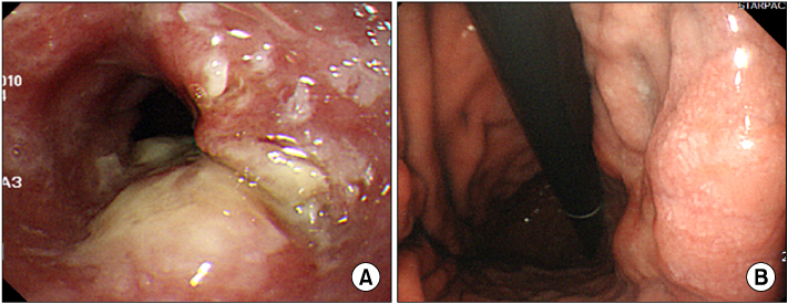
Endoscopic findings. (A) There is a fungating mass at mid-esophagus at 30~35 cm from the incisor. (B) A flat elevated lesion with central depression of lesser curvature from the angle to the high body is noted.
Fig. 2
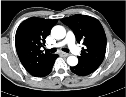
Chest computed tomography findings. The circumferential wall thickening is noted at esophagus, about 3 cm in length, below the carina.
A Case of Double Primary Cancers in the Esophagus and Stomach
Fig. 1
Endoscopic findings. (A) There is a fungating mass at mid-esophagus at 30~35 cm from the incisor. (B) A flat elevated lesion with central depression of lesser curvature from the angle to the high body is noted.
Fig. 2
Chest computed tomography findings. The circumferential wall thickening is noted at esophagus, about 3 cm in length, below the carina.