• Contact us
  • E-Submission
ABOUT
BROWSE ARTICLES
JOURNAL POLICIES
FOR CONTRIBUTORS

Articles

Page Path

Images and Solution

Retrorectal Teratoma with Ovarian Teratoma

The Ewha Medical Journal 2012;35(2):140-142. Published online: September 30, 2012

Department of Surgery, Ewha Womans University School of Medicine, Seoul, Korea.

1Department of Pathology, Ewha Womans University School of Medicine, Seoul, Korea.

2Department of Obstetrics and Gynecology, Ewha Womans University School of Medicine, Seoul, Korea.

Corresponding author: Ryung-Ah Lee, Department of Surgery, Ewha Womans University School of Medicine, 1071 Anyangcheon-ro, Yangcheon-gu, Seoul 158-710, Korea. Tel: 82-2-2650-2659, Fax: 82-2-2644-7984, ralee@ewha.ac.kr
• Received: June 11, 2012   • Accepted: July 16, 2012

Copyright © 2012. Ewha Womans University School of Medicine

This is an Open Access article distributed under the terms of the Creative Commons Attribution Non-Commercial License (http://creativecommons.org/licenses/by-nc/3.0/) which permits unrestricted non-commercial use, distribution, and reproduction in any medium, provided the original work is properly cited.
  • 36 Views
  • 0 Download
prev next
  • 1. Terada T. Mature teratoma of the rectum. Gastrointest Endosc 2010;71:1068-1069.
  • 2. Kaeser MA, McDonald JK, Kettner NW. A calcific pelvic mass in a woman with chronic spinal pain: a case of mature cystic teratoma. J Chiropr Med 2011;10:327-332.
  • 3. Baek EH, Kim KI, Lee AR, Gang G, Kwon YS. Periappendiceal mature cystic teratoma successfully treated with laparoendoscopic surgery. Am Surg 2012;78:70-71.
  • 4. Moawad NS, Starks D, Ashby K. Ectopic ovarian teratoma of the uterosacral ligament associated with a large ovarian dermoid. J Minim Invasive Gynecol 2008;15:523-524.
  • 5. Khoo CK, Chua I, Siow A, Chern B. Parasitic dermoid cyst of the pouch of Douglas: a case report. J Minim Invasive Gynecol 2008;15:761-763.
  • 6. Kahraman K, Kurtay G, Kiremitci S. A presacral dermoid cyst with extremely high serum CA19-9 level. J Obstet Gynaecol 2012;32:103-104.
  • 7. La Quaglia MP, Feins N, Eraklis A, Hendren WH. Rectal duplications. J Pediatr Surg 1990;25:980-984.
  • 8. Nishie A, Yoshimitsu K, Honda H, Irie H, Aibe H, Shinozaki K, et al. Presacral dermoid cyst with scanty fat component: usefulness of chemical shift and diffusion-weighted MR imaging. Comput Med Imaging Graph 2003;27:293-296.
  • 9. Dahan H, Arrivé L, Wendum D, Docou le Pointe H, Djouhri H, Tubiana JM. Retrorectal developmental cysts in adults: clinical and radiologic-histopathologic review, differential diagnosis, and treatment. Radiographics 2001;21:575-584.
  • 10. Guillem P, Ernst O, Herjean M, Triboulet JP. Retrorectal tumors: an assessment of the abdominal approach. Ann Chir 2001;126:138-142.
Fig. 1
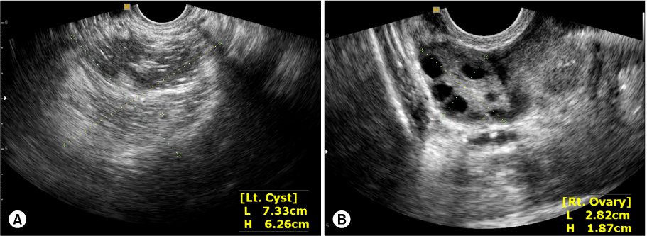
Pelvic ultrasonography findings. (A) 7.3×6.2 cm sized left ovary with inhomogeneous cystic lesion is noted. (B) Normal sized right ovary is demonstrated.
emj-35-140-g001.jpg
Fig. 2
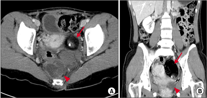
Abdomen & pelvis computed tomography findings. CT images show 5.2×5.8 cm sized cystic mass (indicated by arrow) in the left ovary and 5.5×6.3 cm sized mass (indicated by arrowhead) composed of calcification, fat and soft tissue adjacent to rectum in the presacral area. (A) Axial view. (B) Coronal view.
emj-35-140-g002.jpg
Fig. 3
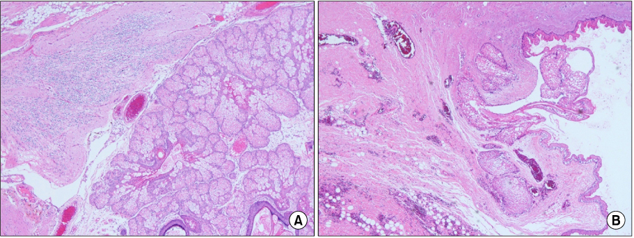
Microscopic findings. Histological findings of the resected specimen in left ovarian mass (A) and retroporitoneal mass (B) are composed of keratinizing stratified squamous epithelium associated with the hair and mucus, which are compatible with mature cystic teratoma (H&E stain, ×40).
emj-35-140-g003.jpg

Figure & Data

References

    Citations

    Citations to this article as recorded by  

      Download Citation

      Download a citation file in RIS format that can be imported by all major citation management software, including EndNote, ProCite, RefWorks, and Reference Manager.

      Format:

      Include:

      Retrorectal Teratoma with Ovarian Teratoma
      Ewha Med J. 2012;35(2):140-142.   Published online September 30, 2012
      Download Citation
      Download a citation file in RIS format that can be imported by all major citation management software, including EndNote, ProCite, RefWorks, and Reference Manager.

      Format:
      • RIS — For EndNote, ProCite, RefWorks, and most other reference management software
      • BibTeX — For JabRef, BibDesk, and other BibTeX-specific software
      Include:
      • Citation for the content below
      Retrorectal Teratoma with Ovarian Teratoma
      Ewha Med J. 2012;35(2):140-142.   Published online September 30, 2012
      Close

      Figure

      • 0
      • 1
      • 2
      Retrorectal Teratoma with Ovarian Teratoma
      Image Image Image
      Fig. 1 Pelvic ultrasonography findings. (A) 7.3×6.2 cm sized left ovary with inhomogeneous cystic lesion is noted. (B) Normal sized right ovary is demonstrated.
      Fig. 2 Abdomen & pelvis computed tomography findings. CT images show 5.2×5.8 cm sized cystic mass (indicated by arrow) in the left ovary and 5.5×6.3 cm sized mass (indicated by arrowhead) composed of calcification, fat and soft tissue adjacent to rectum in the presacral area. (A) Axial view. (B) Coronal view.
      Fig. 3 Microscopic findings. Histological findings of the resected specimen in left ovarian mass (A) and retroporitoneal mass (B) are composed of keratinizing stratified squamous epithelium associated with the hair and mucus, which are compatible with mature cystic teratoma (H&E stain, ×40).
      Retrorectal Teratoma with Ovarian Teratoma
      TOP