1Department of Implant Dentistry, Graduate School of Clinical Dentistry, Ewha Womans University, Seoul, Korea.
2Division of Oral and Maxillofacial Surgery, Department of Dentistry, Ewha Womans University School of Medicine, Seoul, Korea.
3Division of Conservative Dentistry, Department of Dentistry, Ewha Womans University School of Medicine, Seoul, Korea.
Copyright © 2012. Ewha Womans University School of Medicine
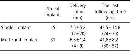
The time of delivery and the last follow-up
Values are presented as mean±SD (range).
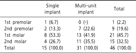
Number of implants in the posterior maxilla
Values are presented as no (%).
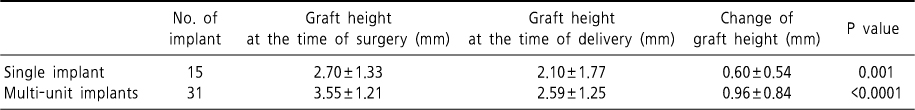
Graft height at the time of surgery and delivery
Values are presented as mean±SD.
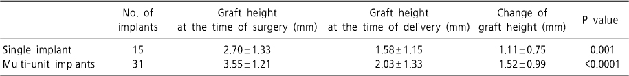
Graft height at the time of surgery and last follow-up
Values are presented as mean±SD.
Values are presented as mean±SD (range).
Values are presented as no (%).
Values are presented as mean±SD.
Values are presented as mean±SD.