Department of Surgery, Ewha Womans University School of Medicine, Seoul, Korea.
1Department of Pathology, Ewha Womans University School of Medicine, Seoul, Korea.
Corresponding author: Ryung-Ah Lee, Department of Surgery, Ewha Womans University School of Medicine, 1071 Anyangcheon-ro, Yangcheon-gu, Seoul 158-710, Korea. Tel: 82-2-2650-2659, Fax: 82-2-2644-7984, ralee@ewha.ac.kr
• Received: August 23, 2011 • Accepted: October 28, 2011
Desmoid tumor is rare neoplasm originated from fibrous sheath or musculoaponeurotic structure. It is classified as benign tumor histologically, but clinically, it has malignant characteristics due to its infiltrative growth to adjacent organ and frequent local recurrence. Especially, mesenteric desmoid tumor shows poor prognosis because of its symptoms of pain, intestinal obstruction, ureter obstruction and fistula formation and high frequency of recurrence. We experienced a case of mesenteric desmoid tumor in a 64-year-old woman with a painless abdominal mass. Laparoscopic exploration was performed and 10 cm sized mesenteric mass was identified, which resected widely and the diagnosis was confirmed with desmoid tumor by pathologic report. We reviewed the feature of the mesenteric desmoid tumor, that is, pathophysiology, clinical presentations, diagnosis, treatment and prognosis.
2. Kawashima A, Goldman SM, Fishman EK, Kuhlman JE, Onitsuka H, Fukuya T, et al. CT of intraabdominal desmoid tumors: is the tumor different in patients with Gardner's disease? AJR Am J Roentgenol 1994;162:339-342.
3. Park BH, Kim HJ, Chang YW, Kim KJ, Lee DK, Dong SH, et al. Desmoid tumor and duodenal adenoma in a patient with familial adenomatous polyposis: a case report. Korean J Gastrointest Endosc 2001;23:32-35.
4. Lee HS, Jeon HM, Ok ST, Kim JS, Lee EJ, Kim JS. Unresectable desmoid tumor developing after surgery of F.A.P case report. J Korean Soc Coloproctol 1998;14:323-329.
5. Shiu MH, Weinstein L, Hajdu SI, Brennan MF. Malignant soft-tissue tumors of the anterior abdominal wall. Am J Surg 1989;158:446-451.
9. Lee JC, Thomas JM, Phillips S, Fisher C, Moskovic E. Aggressive fibromatosis: MRI features with pathologic correlation. AJR Am J Roentgenol 2006;186:247-254.
10. Klein WA, Miller HH, Anderson M, DeCosse JJ. The use of indomethacin, sulindac, and tamoxifen for the treatment of desmoid tumors associated with familial polyposis. Cancer 1987;60:2863-2868.
11. Murayama T, Imoto S, Ito M, Matsushita K, Matozaki S, Nakagawa T, et al. Mesenteric fibromatosis presenting as fever of unknown origin. Am J Gastroenterol 1992;87:1503-1505.
12. Yu YH, Son BK, Jun DW, Kim SH, Jo YJ, Park YS, et al. A case of desmoid tumor presenting as intra-abdominal abscess. Korean J Gastroenterol 2009;53:315-319.
17. Monihan JM, Carr NJ, Sobin LH. CD34 immunoexpression in stromal tumours of the gastrointestinal tract and in mesenteric fibromatoses. Histopathology 1994;25:469-473.
18. Montgomery E, Torbenson MS, Kaushal M, Fisher C, Abraham SC. Beta-catenin immunohistochemistry separates mesenteric fibromatosis from gastrointestinal stro al tumor and sclerosing mesenteritis. Am J Surg Pathol 2002;26:1296-1301.
19. Nam KH, Kweon BC, Lee HK, Lee DW, Woo CK, Park JS, et al. A case of mesenteric fibromatosis after appendectomy. Korean J Med 1998;54:577-581.
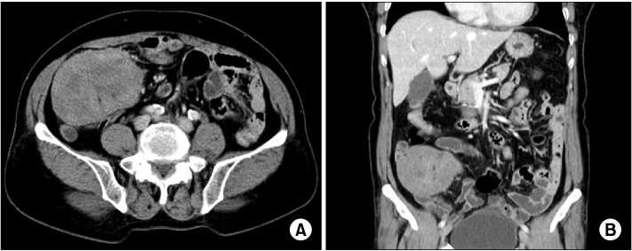
Fig. 1
Abdominal CT findings. 10×6.5 cm sized solid mass is visible in right lower quadrant. The tumor originates from small bowel mesentery. (A) Horizontal view. (B) Coronal view.
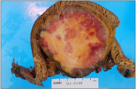
Fig. 2
Gross pathologic finding. 9.5×9×5 cm sized well demarcated mass is identified in the small bowel mesentery. Retraction diverticulum is visible on ileal mucosa.
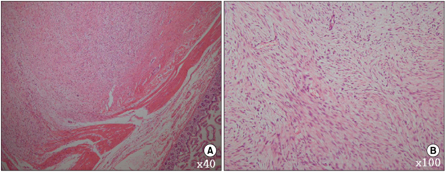
Fig. 3
Micropathologic finding. (A) The tumor infiltrates into the muscle layers of small bowel (H&E stain, ×40). (B) Fibroblastic spindle cells are arranged orderly in collagenous or myxoid matrix (H&E stain, ×100).
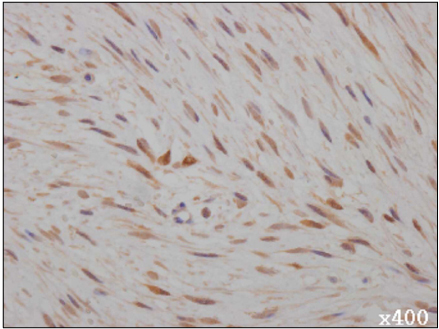
Fig. 4
Immunohistochemical stain for β-catenin shows nuclear and cytoplasmic positivity in tumor cells (Immunohistochemical stain, ×400).
A Case of Ileal Mesenteric Desmoid Tumor Resected by Laparoscopic Surgery
Fig. 1
Abdominal CT findings. 10×6.5 cm sized solid mass is visible in right lower quadrant. The tumor originates from small bowel mesentery. (A) Horizontal view. (B) Coronal view.
Fig. 2
Gross pathologic finding. 9.5×9×5 cm sized well demarcated mass is identified in the small bowel mesentery. Retraction diverticulum is visible on ileal mucosa.
Fig. 3
Micropathologic finding. (A) The tumor infiltrates into the muscle layers of small bowel (H&E stain, ×40). (B) Fibroblastic spindle cells are arranged orderly in collagenous or myxoid matrix (H&E stain, ×100).
Fig. 4
Immunohistochemical stain for β-catenin shows nuclear and cytoplasmic positivity in tumor cells (Immunohistochemical stain, ×400).
Fig. 1
Fig. 2
Fig. 3
Fig. 4
A Case of Ileal Mesenteric Desmoid Tumor Resected by Laparoscopic Surgery