Department of Surgery, Ewha Womans University School of Medicine, Seoul, Korea.
1Department of Pathology, Ewha Womans University School of Medicine, Seoul, Korea.
Corresponding author: Ryung-Ah Lee, Department of Surgery, Ewha Womans University School of Medicine, 1071 Anyangcheon-ro, Yangcheon-gu, Seoul 158-710, Korea. Tel: 82-2-2650-2659, Fax: 82-2-2644-7984, ralee@ewha.ac.kr
• Received: February 9, 2012 • Accepted: February 28, 2012
1. Bardaji M, Roset F, Camps R, Sant F, Fernandez-Layos MJ. Symptomatic colonic lipoma: differential diagnosis of large bowel tumors. Int J Colorectal Dis 1998;13:1-2.
4. Kim BC, Jung SW, Kwon SH, Park JS, Ko BK, Kim YM, et al. A case of jejuno-jejunal intussusception caused by a small intestinal lipoma. Korean J Med 2008;75:333-336.
5. Pemberton LB, Manax WG. Complete obstruction of the colon by lipoma. Surgery 1971;69:139-141.
6. Ryu KW, Kim DS, Hong BW, Lee JB, Moon HY, Choi SY. Diagnosis and treatment of adult intussusception due to gastrointestinal lipoma. J Korean Surg Soc 2000;59:61-66.
7. Heiken JP, Forde KA, Gold RP. Computed tomography as a definitive method for diagnosing gastrointestinal lipomas. Radiology 1982;142:409-414.
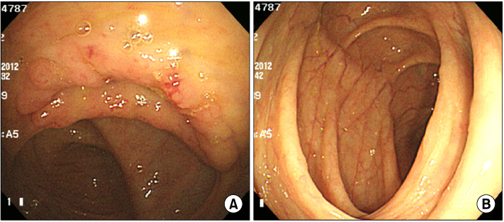
Colonoscopic findings. (A) Hyperemic ileocecal valve was observed. (B) No visible mass is found in the hepatic flexure of ascending colon.
Fig. 2
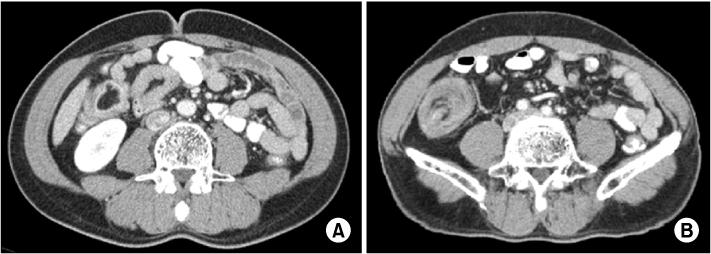
Abdominal computed tomography findings. (A) Well-defined, 3 cm sized low attenuated submucosal mass is identified on the hepatic flexure of ascending colon. (B) A round target-shaped mass is revealed in the right lower quadrant consisting of different densities.
Fig. 3
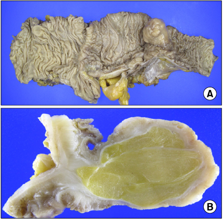
Macroscopic findings. (A) An ovoid shaped pedunculated submucosal mass is observed. (B) The cut surface shows lobulated yellowish adipose tissue with mixed thin fibrous tissue.
Fig. 4
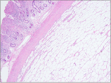
Microscopic finding. The mass is located in the submucosa and composed of mature lipocytes (H&E stain, ×40).
A Case of Ileal Lipoma Misdiagnosed as Colonic Lipoma
Fig. 1
Colonoscopic findings. (A) Hyperemic ileocecal valve was observed. (B) No visible mass is found in the hepatic flexure of ascending colon.
Fig. 2
Abdominal computed tomography findings. (A) Well-defined, 3 cm sized low attenuated submucosal mass is identified on the hepatic flexure of ascending colon. (B) A round target-shaped mass is revealed in the right lower quadrant consisting of different densities.
Fig. 3
Macroscopic findings. (A) An ovoid shaped pedunculated submucosal mass is observed. (B) The cut surface shows lobulated yellowish adipose tissue with mixed thin fibrous tissue.
Fig. 4
Microscopic finding. The mass is located in the submucosa and composed of mature lipocytes (H&E stain, ×40).
Fig. 1
Fig. 2
Fig. 3
Fig. 4
A Case of Ileal Lipoma Misdiagnosed as Colonic Lipoma