• Contact us
  • E-Submission
ABOUT
BROWSE ARTICLES
JOURNAL POLICIES
FOR CONTRIBUTORS

Articles

Case Report

Clostridium difficile Colitis after Topical Ophthalmic Use of Levofloxacin

The Ewha Medical Journal 2013;36(Suppl):S5-S8. Published online: December 23, 2013

Department of Internal Medicine, Daerim Saint Mary's Hospital, Seoul, Korea.

Corresponding author: Seong Yong Woo. Department of Internal Medicine, Daerim Saint Mary's Hospital, 657, Siheung-daero, Yeongdeungpo-gu, Seoul 150-822, Korea. Tel: 82-2-829-9301, Fax: 82-2-829-9299, loveandy@hanmail.net
• Received: June 12, 2013   • Accepted: July 2, 2013

Copyright © 2013. Ewha Womans University School of Medicine

This is an Open Access article distributed under the terms of the Creative Commons Attribution Non-Commercial License (http://creativecommons.org/licenses/by-nc/3.0/) which permits unrestricted non-commercial use, distribution, and reproduction in any medium, provided the original work is properly cited.
  • 55 Views
  • 0 Download
  • 2 Crossref
prev
  • Clostridium difficile colitis (CDC) has been associated with virtually all available antibiotics. It is well known that administration of drugs by the ocular route can result in systemic absorption with the possibility of systemic effect. We report a case of a 74-year-old woman who was treated with 0.5% levofloxacin eyedrops for ten weeks running for the post operative management of two separate bilateral cataract surgeries. Toxin assay for C. difficile was positive and the endoscopic findings were compatible with CDC. The plasma concentrations reached after intraocular administration of levofloxacin seem so trivial, but the administration of topical agents could make accumulated effect and be a potential cause of systemic toxicity.
  • 1. Larson HE, Price AB, Honour P, Borriello SP. Clostridium difficile and the aetiology of pseudomembranous colitis. Lancet 1978;1:1063-1066.
  • 2. Fraunfelder FT, Bagby GC Jr, Kelly DJ. Fatal aplastic anemia following topical administration of ophthalmic chloramphenicol. Am J Ophthalmol 1982;93:356-360.
  • 3. Coleman AL, Diehl DL, Jampel HD, Bachorik PS, Quigley HA. Topical timolol decreases plasma high-density lipoprotein cholesterol level. Arch Ophthalmol 1990;108:1260-1263.
  • 4. Bowman RJ, Cope J, Nischal KK. Ocular and systemic side effects of brimonidine 0.2% eye drops (Alphagan) in children. Eye (Lond) 2004;18:24-26.
  • 5. Centers for Disease Control and Prevention (CDC)Severe Clostridium difficile-associated disease in populations previously at low risk: four states, 2005. MMWR Morb Mortal Wkly Rep 2005;54:1201-1205.
  • 6. Cohen SH, Gerding DN, Johnson S, Kelly CP, Loo VG, McDonald LC, et al. Clinical practice guidelines for Clostridium difficile infection in adults: 2010 update by the society for healthcare epidemiology of America (SHEA) and the infectious diseases society of America (IDSA). Infect Control Hosp Epidemiol 2010;31:431-455.
  • 7. Muto CA, Pokrywka M, Shutt K, Mendelsohn AB, Nouri K, Posey K, et al. A large outbreak of Clostridium difficile-associated disease with an unexpected proportion of deaths and colectomies at a teaching hospital following increased fluoroquinolone use. Infect Control Hosp Epidemiol 2005;26:273-280.
  • 8. Vistakon Pharmaceuticals LLCDailyMed: Quixin (levofloxacin) solution [Internet] Bethesda (MD), National Institutes of Health, Health & Human Services. 2011;cited 2013 Mar. Available from: http://dailymed.nlm.nih.gov/dailymed/lookup.cfm?setid=09b3a912-97f9-45ae-a162-9f45c860dc22
  • 9. Gaynor BD, Chidambaram JD, Cevallos V, Miao Y, Miller K, Jha HC, et al. Topical ocular antibiotics induce bacterial resistance at extraocular sites. Br J Ophthalmol 2005;89:1097-1099.
  • 10. Nelson WL, Fraunfelder FT, Sills JM, Arrowsmith JB, Kuritsky JN. Adverse respiratory and cardiovascular events attributed to timolol ophthalmic solution, 1978-1985. Am J Ophthalmol 1986;102:606-611.
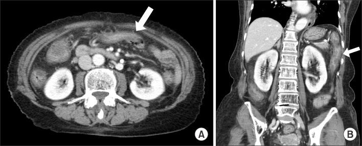
Fig. 1
Abdominal computed tomography findings. They show diffuse edematous wall thickenings (white arrows) of the entire colon (A, B).
emj-36-S5-g001.jpg
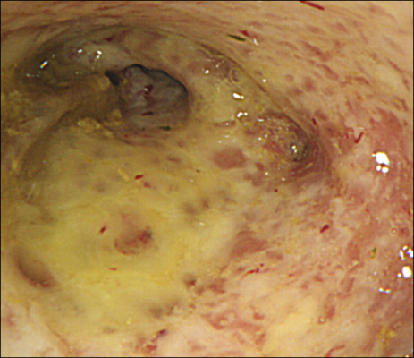
Fig. 2
Sigmoidoscopic finding. It shows multiple yellowish plaques with erythematous and edematous mucosa in the sigmoid colon.
emj-36-S5-g002.jpg
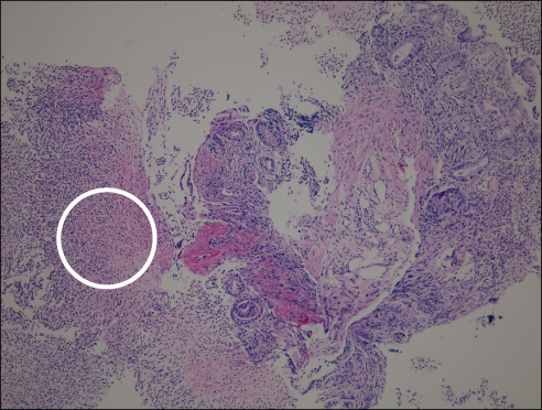
Fig. 3
Pathologic finding of biopsy specimen from colon mucosa. It shows severe erosion with pseudomembrane composed of fibrinous neutrophilic exudates (white circle) (H&E, ×100).
emj-36-S5-g003.jpg
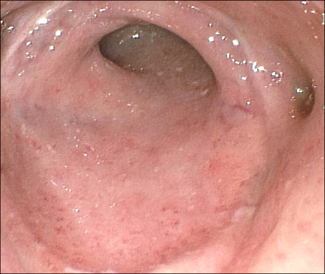
Fig. 4
Follow-up endoscopic finding. It shows a nearly improved state of colitis with no pseudomembrane.
emj-36-S5-g004.jpg

Figure & Data

References

    Citations

    Citations to this article as recorded by  
    • Concurrent Use of Sulfonylureas and Antimicrobials of the Elderly in Korea: A Potential Risk of Hypoglycemia
      Sera Lee, Miyoung Ock, Hyunah Kim
      Korean Journal of Clinical Pharmacy.2018; 28(3): 188.     CrossRef
    • Levofloxacin

      Reactions Weekly.2014; 1490(1): 25.     CrossRef

    Download Citation

    Download a citation file in RIS format that can be imported by all major citation management software, including EndNote, ProCite, RefWorks, and Reference Manager.

    Format:

    Include:

    Clostridium difficile Colitis after Topical Ophthalmic Use of Levofloxacin
    Ewha Med J. 2013;36(Suppl):S5-S8.   Published online December 23, 2013
    Download Citation
    Download a citation file in RIS format that can be imported by all major citation management software, including EndNote, ProCite, RefWorks, and Reference Manager.

    Format:
    • RIS — For EndNote, ProCite, RefWorks, and most other reference management software
    • BibTeX — For JabRef, BibDesk, and other BibTeX-specific software
    Include:
    • Citation for the content below
    Clostridium difficile Colitis after Topical Ophthalmic Use of Levofloxacin
    Ewha Med J. 2013;36(Suppl):S5-S8.   Published online December 23, 2013
    Close

    Figure

    • 0
    • 1
    • 2
    • 3
    Clostridium difficile Colitis after Topical Ophthalmic Use of Levofloxacin
    Image Image Image Image
    Fig. 1 Abdominal computed tomography findings. They show diffuse edematous wall thickenings (white arrows) of the entire colon (A, B).
    Fig. 2 Sigmoidoscopic finding. It shows multiple yellowish plaques with erythematous and edematous mucosa in the sigmoid colon.
    Fig. 3 Pathologic finding of biopsy specimen from colon mucosa. It shows severe erosion with pseudomembrane composed of fibrinous neutrophilic exudates (white circle) (H&E, ×100).
    Fig. 4 Follow-up endoscopic finding. It shows a nearly improved state of colitis with no pseudomembrane.
    Clostridium difficile Colitis after Topical Ophthalmic Use of Levofloxacin
    TOP