This is an Open Access article distributed under the terms of the Creative Commons Attribution Non-Commercial License (http://creativecommons.org/licenses/by-nc/3.0/) which permits unrestricted non-commercial use, distribution, and reproduction in any medium, provided the original work is properly cited.
Intraductal tubulopapillary neoplasm (ITPN) of the pancreas has been recently reported. It is very rare, therefore clinical behavior and prognosis has not yet been characterized. We experienced a case of ITPN of the pancreas which presented with acute pancreatitis and treated with Whipple's operation. Histopathologic finding showed papillary hyperplasia with carcinomatous change. The tumor recurred after 47 month of operation, and she underwent total pancreatectomy. Pathologic finding revealed tubulopapillary growth with high grade dysplasia. Immunohistochemial staining was not performed, however gross and microscopic findings were compatible with ITPN of the pancreas. We report a case of ITPN of the pancreas.
1. Yamaguchi H, Shimizu M, Ban S, Koyama I, Hatori T, Fujita I, et al. Intraductal tubulopapillary neoplasms of the pancreas distinct from pancreatic intraepithelial neoplasia and intraductal papillary mucinous neoplasms. Am J Surg Pathol 2009;33:1164-1172.
2. Longnecker DS, Adler G, Hruban RH, Kloppel G. Intraductal papillary-mucinous neoplasms of the pancreas. In: Hamilton SR, Aaltonen LA, editors. Pathology and genetics of tumours of the digestive system WHO classification of tumours. Lyon, France: IARC Press; 2000. p. 237-240.
3. Bosman FT, Carneiro F, Hruban RH, Theise ND. WHO classification of tumours of the digestive system. 2nd ed. Lyon, France: IARC Press; 2010.
4. Bhuva N, Wasan H, Spalding D, Stamp G, Harrison M. Intraductal tubulopapillary neoplasm of the pancreas as a radiation induced malignancy. BMJ Case Rep 2011;2011:pii: bcr0920114777. 10.1136/bcr.09.2011.4777
5. Guan H, Gurda G, Marie Lennon A, Hruban RH, Erozan YS. Intraductal tubulopapillary neoplasm of the pancreas on fine needle aspiration: case report with differential diagnosis. Diagn Cytopathol 2012 07 16 [Epub]. 10.1002/dc.22890
6. Jokoji R, Tsuji H, Tsujimoto M, Shinno N, Tori M. Intraductal tubulopapillary neoplasm of pancreas with stromal osseous and cartilaginous metaplasia; a case report. Pathol Int 2012;62:339-343.
8. Hruban RH, Adsay NV, Albores-Saavedra J, Compton C, Garrett ES, Goodman SN, et al. Pancreatic intraepithelial neoplasia: a new nomenclature and classification system for pancreatic duct lesions. Am J Surg Pathol 2001;25:579-586.
9. Furukawa T, Kloppel G, Volkan Adsay N, Albores-Saavedra J, Fukushima N, Horii A, et al. Classification of types of intraductal papillary-mucinous neoplasm of the pancreas: a consensus study. Virchows Arch 2005;447:794-799.
11. Park HJ, Jang KT, Heo JS, Choi YL, Han J, Kim SH. A potential case of intraductal tubulopapillary neoplasms of the bile duct. Pathol Int 2010;60:630-635.
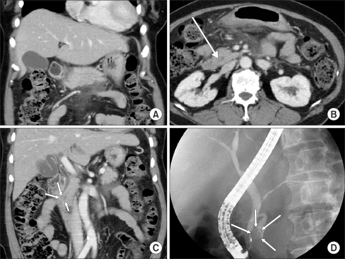
Abdomen computed tomography (CT) findings. (A) Diffuse parenchymal swellling of pancreas, peripancreatic fatty infiltration and fluid collection are seen. (B, C) A small nodule and scanty dilatation of pancreatic duct in the pancreas head are seen on the CT scan (arrows). (D) Endoscopic retrograde cholangiopancreatography findings. A nodular filling defect and duct dilatation of pancreatic head portion and common bile duct dilatation are seen on the cholangiopancreatogram (arrows).
Fig. 2
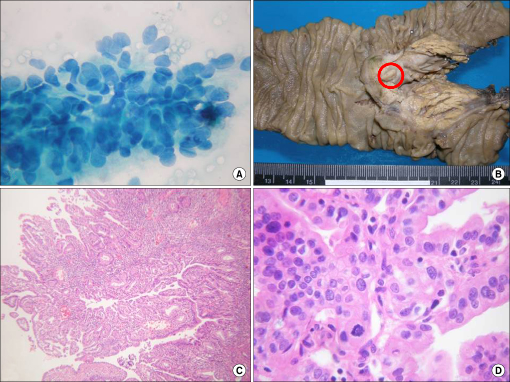
Histopathologic findings. (A) Brush cytology picture shows hyperchromatic nucleus and nuclear molding, therefore those are suspected to be malignant cells. (B) Gross finding after resection shows a 1.6×1.1 cm sized polypoid mass (red circle) is seen on the proximal portion of dilated pancreatic duct. (C) Tubulopapillary proliferations are seen (H&E, ×40). (D) Cells with nuclear polymorphism, hyperchromatism, and mitosis are seen, therefore high grade dysplasia or carcinomatous change is suspected (H&E, ×250).
Fig. 3
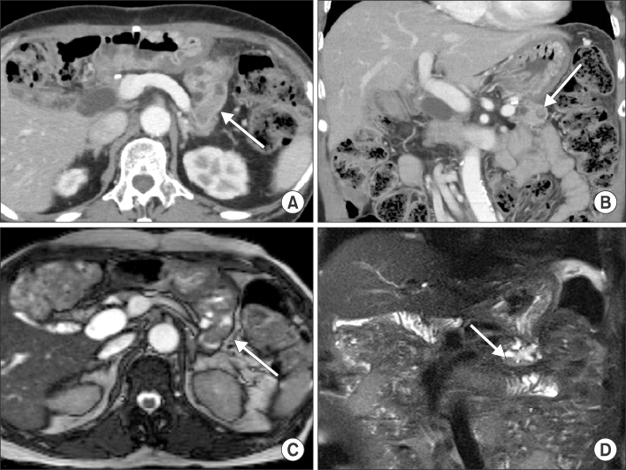
(A, B) Computed tomography (CT) findings after 47 months of resection. Axial and coronal CT scan shows diffuse pancreatic duct dilatation with enhancing mass (arrow) inside of the duct. (C, D) Magnetic resonance imaging findings after 47 month of resection. T2 weighted images also shows mass like lesion (arrow) inside of dilated pancreatic duct after 47 months of resection.
Fig. 4
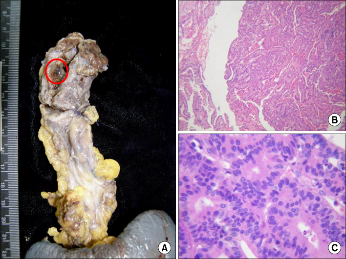
(A) Gross sample of 2nd operation (total pancreatectomy and splenectomy). A brownish mass like lesion mixed with brownish sludge material (red circle) is seen inside of dilated pancreatic duct. (B) Tubulopapillary proliferation is seen (H&E, ×40). (C) Nuclear polymorphism and hyperchomatic nucleus suggests high grade dysplasia (H&E, ×250).
Acute Pancreatitis Associated with Intraductal Tubulopapillary Neoplasm of the Pancreas
Fig. 1
Abdomen computed tomography (CT) findings. (A) Diffuse parenchymal swellling of pancreas, peripancreatic fatty infiltration and fluid collection are seen. (B, C) A small nodule and scanty dilatation of pancreatic duct in the pancreas head are seen on the CT scan (arrows). (D) Endoscopic retrograde cholangiopancreatography findings. A nodular filling defect and duct dilatation of pancreatic head portion and common bile duct dilatation are seen on the cholangiopancreatogram (arrows).
Fig. 2
Histopathologic findings. (A) Brush cytology picture shows hyperchromatic nucleus and nuclear molding, therefore those are suspected to be malignant cells. (B) Gross finding after resection shows a 1.6×1.1 cm sized polypoid mass (red circle) is seen on the proximal portion of dilated pancreatic duct. (C) Tubulopapillary proliferations are seen (H&E, ×40). (D) Cells with nuclear polymorphism, hyperchromatism, and mitosis are seen, therefore high grade dysplasia or carcinomatous change is suspected (H&E, ×250).
Fig. 3
(A, B) Computed tomography (CT) findings after 47 months of resection. Axial and coronal CT scan shows diffuse pancreatic duct dilatation with enhancing mass (arrow) inside of the duct. (C, D) Magnetic resonance imaging findings after 47 month of resection. T2 weighted images also shows mass like lesion (arrow) inside of dilated pancreatic duct after 47 months of resection.
Fig. 4
(A) Gross sample of 2nd operation (total pancreatectomy and splenectomy). A brownish mass like lesion mixed with brownish sludge material (red circle) is seen inside of dilated pancreatic duct. (B) Tubulopapillary proliferation is seen (H&E, ×40). (C) Nuclear polymorphism and hyperchomatic nucleus suggests high grade dysplasia (H&E, ×250).
Fig. 1
Fig. 2
Fig. 3
Fig. 4
Acute Pancreatitis Associated with Intraductal Tubulopapillary Neoplasm of the Pancreas