• Contact us
  • E-Submission
ABOUT
BROWSE ARTICLES
JOURNAL POLICIES
FOR CONTRIBUTORS

Articles

Page Path

Case Report

Serosal Type Eosinophilic Gastroenteritis

The Ewha Medical Journal 2014;37(2):126-130. Published online: September 30, 2014

Department of Internal Medicine, Ewha Womans University College of Medicine, Seoul, Korea.

Corresponding author: Tae Hun Kim. Department of Internal Medicine, Ewha Womans University Mokdong Hospital, 1071 Anyangcheon-ro, Yangcheon-gu, Seoul 158-710, Korea. Tel: 82-2-2650-2724, Fax: 82-2-2655-2076, thkm@ewha.ac.kr
• Received: March 16, 2014   • Accepted: June 26, 2014

Copyright © 2014. Ewha Womans University School of Medicine

This is an Open Access article distributed under the terms of the Creative Commons Attribution Non-Commercial License (http://creativecommons.org/licenses/by-nc/3.0/) which permits unrestricted non-commercial use, distribution, and reproduction in any medium, provided the original work is properly cited.
  • 70 Views
  • 0 Download
  • 1 Crossref
prev next
  • Eosinophilic gastroenteritis is a rare, benign condition, characterized by various gastrointestinal symptoms associated with eosinophilic infiltration of the wall of the any part of the digestive tract, most commonly the stomach and small intestine. Eosinophilic gastroenteritis is generally classified according to the involved layer of the gastrointestinal tract. Serosal type is the rarest form of eosinophilic gastroenteritis that is characteristically accompanied with eosinophilic ascites and responds well to steroid treatment. We have experienced a typical case of serosal type eosinophilic gastroenteritis in a women who complained of abdominal pain. She had peripheral eosinophilia, gastric and small bowel wall thickening with eosionophilic ascites. Her symptom relieved rapidly after starting corticosteroid treatment and she had long been in clinical remission after discontinuation of corticosteroid administration.
  • 1. Mendez-Sanchez N, Chavez-Tapia NC, Vazquez-Elizondo G, Uribe M. Eosinophilic gastroenteritis: a review. Dig Dis Sci 2007;52:2904-2911.
  • 2. Baig MA, Qadir A, Rasheed J. A review of eosinophilic gastroenteritis. J Natl Med Assoc 2006;98:1616-1619.
  • 3. Kaijser R. Allergic disease of the gut from the point of view of the surgeon. Arch Klin Chir 1937;188:36-64.
  • 4. Ureles AL, Alschibaja T, Lodico D, Stabins SJ. Idiopathic eosinophilic infiltration of the gastrointestinal tract, diffuse and circumscribed: a proposed classification and reiew of the literature, with two additional cases. Am J Med 1961;30:899-909.
  • 5. Klein NC, Hargrove RL, Sleisenger MH, Jeffries GH. Eosinophilic gastroenteritis. Medicine (Baltimore) 1970;49:299-319.
  • 6. Kim JD, Im EH, Lee JH, Lee TH, Kim SM, Choi YW, et al. A case of eosinophilic esophagogastroenteritis with transmural involvement. Korean J Gastrointest Endosc 2007;35:404-409.
  • 7. Kim NI, Jo YJ, Song MH, Kim SH, Kim TH, Park YS, et al. Clinical features of eosinophilic gastroenteritis. Korean J Gastroenterol 2004;44:217-223.
  • 8. Lee SJ, Chung WC, Lee KM, Lee BI, Kim YC, Hong CK, et al. A case of eosinophilic gastroenteritis in a patient with traumatic hypopituitarism. Korean J Gastrointest Endosc 2007;34:51-55.
  • 9. Na YJ, Shim KN, Yeom MS, Kim HS, Jung SA, Yoo K, et al. A case of eosinophilic gastroenteritis with eosinophilic ascites. Korean J Gastrointest Endosc 2004;29:22-26.
  • 10. Kim TG, Park J, Seo EH, Joo HR, Park SH, Kim TO, et al. Esosinophilic gastroenteritis with clostridium difficile-associated colitis: a case report. Korean J Gastrointest Endosc 2011;43:64-68.
  • 11. Jung YJ, Kim MH, Lee SY, Kim MK, Lee SS, Lee SK, et al. A case of eosinophilic gastroenteritis associated with eosinophilic cholecystitis. Korean J Med 2005;69:Suppl 3. S758-S762.
  • 12. Kim SS, Choi CH, Choi HS, Park IW, Gham CW, Cho HG, et al. A case of eosinophilic enterocolitis associated with eosinophilic ascites and cystitis. Korean J Med 2005;69:Suppl 3. S746-S752.
  • 13. Hyun JN, Lee JH, Lee JY, Kim SM, Yoo WJ, Kim SY, et al. A case of eosinophilic gastroenteritis with eosinophilic ascites. Korean J Med 2007;73:535-538.
  • 14. Jee SC, Kim HK, Kong KT, Lee YJ, Jung KM, Kim JO, et al. A case of recurred gastric dysplasia associated with eosinophilic gastroenteritis. Korean J Med 2007;73:530-534.
  • 15. Yoon CO, Lee HL, Lee OY, Yoon BC, Choi HS, Hahm JS, et al. A case of eosinophilic gastroenteritis presenting as massive ascites. Korean J Med 2007;72:Suppl 2. S121-S125.
  • 16. Park CH, Lee SJ, Chang HJ, Lee GJ, Lee H, Song HS, et al. A case of eosionophilic gastroenteritis involving entire gastrointestinal tract with eosinophilic ascites. Korean J Gastrointest Endosc 2008;36:282-287.
  • 17. Kang B, Chung WC, Lee KM, Paik CN, Lee JM, Jeon HS, et al. A case of eosinophilic gastroenteritis presenting with fever and multiple lymphadenopathy. Korean J Gastrointest Endosc 2011;42:361-365.
Fig. 1
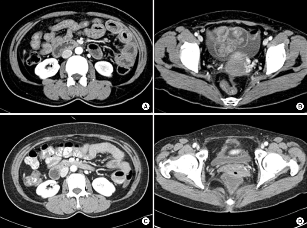
Abdominopelvic computed tomography (CT) scan findings. The initial CT scan shows wall thickening of small bowel (A), and small amount ascites in the pelvic cavity (B). The CT scan after prednisolone treatment shows interval improvement of wall thickening of small bowel (C), and ascites in the pelvic cavity (D).
emj-37-126-g001.jpg
Fig. 2
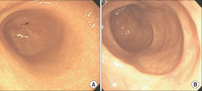
Endoscopic findings. There are non-specific superficial mucosal erythema on esophagogastroscopy (A) and entire colonic mucosa is normal on colonoscopic examination (B).
emj-37-126-g002.jpg
Fig. 3
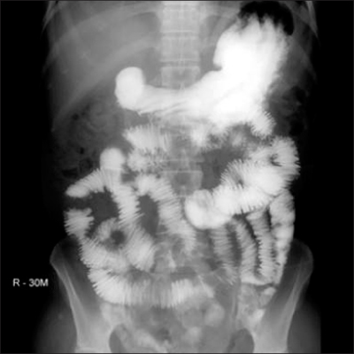
Small bowel series. It shows coin stacked appearance and multifocal fold thickening and thumb printing of nearly entire jejunum.
emj-37-126-g003.jpg
Table 1
Analysis of the reported cases of eosinophilic gastroenteritis last decade
emj-37-126-i001.jpg

Figure & Data

References

    Citations

    Citations to this article as recorded by  
    • Eosinophilic Ascites: A Rare Diagnosis With an Even Rarer Etiology
      Devipriya Surapaneni, Bilal Azam, Sharath Chandra Dasi
      Cureus.2024;[Epub]     CrossRef

    Download Citation

    Download a citation file in RIS format that can be imported by all major citation management software, including EndNote, ProCite, RefWorks, and Reference Manager.

    Format:

    Include:

    Serosal Type Eosinophilic Gastroenteritis
    Ewha Med J. 2014;37(2):126-130.   Published online September 30, 2014
    Download Citation
    Download a citation file in RIS format that can be imported by all major citation management software, including EndNote, ProCite, RefWorks, and Reference Manager.

    Format:
    • RIS — For EndNote, ProCite, RefWorks, and most other reference management software
    • BibTeX — For JabRef, BibDesk, and other BibTeX-specific software
    Include:
    • Citation for the content below
    Serosal Type Eosinophilic Gastroenteritis
    Ewha Med J. 2014;37(2):126-130.   Published online September 30, 2014
    Close

    Figure

    • 0
    • 1
    • 2
    Serosal Type Eosinophilic Gastroenteritis
    Image Image Image
    Fig. 1 Abdominopelvic computed tomography (CT) scan findings. The initial CT scan shows wall thickening of small bowel (A), and small amount ascites in the pelvic cavity (B). The CT scan after prednisolone treatment shows interval improvement of wall thickening of small bowel (C), and ascites in the pelvic cavity (D).
    Fig. 2 Endoscopic findings. There are non-specific superficial mucosal erythema on esophagogastroscopy (A) and entire colonic mucosa is normal on colonoscopic examination (B).
    Fig. 3 Small bowel series. It shows coin stacked appearance and multifocal fold thickening and thumb printing of nearly entire jejunum.
    Serosal Type Eosinophilic Gastroenteritis

    Analysis of the reported cases of eosinophilic gastroenteritis last decade

    Table 1 Analysis of the reported cases of eosinophilic gastroenteritis last decade

    TOP