• Contact us
  • E-Submission
ABOUT
BROWSE ARTICLES
JOURNAL POLICIES
FOR CONTRIBUTORS

Articles

Page Path

Case Report

Toxic Megacolon Associated with Secondary Amyloidosis: An Unusual Complication of Clostridium difficile Colitis

The Ewha Medical Journal 2014;37(1):52-55. Published online: March 25, 2014

Department of Internal Medicine, Ewha Womans University School of Medicine, Seoul, Korea.

Corresponding author: Hye-Kyung Jung. Department of Internal Medicine, Ewha Womans University School of Medicine, 1071 Anyangcheon-ro, Yangcheon-gu, Seoul 158-710, Korea. Tel: 82-2-2650-2874, Fax: 82-2-2655-2874, junghk@ewha.ac.kr
• Received: September 6, 2013   • Accepted: October 18, 2013

Copyright © 2014. Ewha Womans University School of Medicine

This is an Open Access article distributed under the terms of the Creative Commons Attribution Non-Commercial License (http://creativecommons.org/licenses/by-nc/3.0/) which permits unrestricted non-commercial use, distribution, and reproduction in any medium, provided the original work is properly cited.
  • 39 Views
  • 0 Download
prev next
  • Amyloidosis is characterized by extracellular deposition of protein fibrils in one or multiple organs. AA amyloidosis is secondarily occurred to be related with chronic infections or inflammatory diseases. We report a 67-year-old man suffered from secondary AA amyloidosis related with chronic Clostridium difficile colitis after repeated total hip replacement surgery. Infection control is the most important treatment of AA amyloidosis secondary to chronic infection. However, the patient's C. difficile colitis was not controlled well, eventually toxic megacolon with sepsis was developed. Consequently, he had to take total colectomy, but he expired with multi-organ failures. We suggested that early surgical procedure might be one option for intractable C. difficile colitis complicated with secondary amyloidosis.
  • 1. Glenner GG. Amyloid deposits and amyloidosis: the beta-fibril-loses (first of two parts). N Engl J Med 1980;302:1283-1292.
  • 2. Ku JR, Kim A, Hur Y, Suh DW, Park KH, Kim EJ, et al. A case of protein-losing gastroenteropathy due to secondary gastrointestinal amyloidosis in a patient with rheumatoid arthritis. Korean J Med 1997;53:S840-S845.
  • 3. Hunter AM, Borsey DQ, Campbell IW, Macaulay RA. Protein-losing enteropathy due to gastro-intestinal amyloidosis. Postgrad Med J 1979;55:822-823.
  • 4. Lee MH, Park WD, Kim BH, Lee JI, Chang YW, Chang R, et al. Generalized primary amyloidosis with malabsorption syndrome. Korean J Gastrointest Endosc 1987;7:91-96.
  • 5. Kim JS, Kwon SY, Song GG, Shin SW, Kim JS, Bak YT, et al. A case of systemic amyloidosis with intestinal pseudoobstruction. Korean J Gastrointest Endosc 1993;13:375-379.
  • 6. Riemann JF, Schmidt H. Ultrastructural changes in the gut autonomic nervous system following laxative abuse and in other conditions. Scand J Gastroenterol Suppl 1982;71:111-124.
  • 7. Bartlett JG, Moon N, Chang TW, Taylor N, Onderdonk AB. Role of Clostridium difficile in antibiotic-associated pseudomembranous colitis. Gastroenterology 1978;75:778-782.
  • 8. Koh DH, Lee HL, Kim JM, Moon W, Lee OY, Yoon BC, et al. A case of toxic megacolon associated with fulminant pseudomembranous colitis. Korean J Gastrointest Endosc 2008;36:112-116.
  • 9. Redelings MD, Sorvillo F, Mascola L. Increase in Clostridium difficile-related mortality rates, United States, 1999-2004. Emerg Infect Dis 2007;13:1417-1419.
  • 10. Chang J, Rohwer RG. Clostridium difficile infection in adult hamsters. Lab Anim Sci 1991;41:548-552.
  • 11. Longo WE, Mazuski JE, Virgo KS, Lee P, Bahadursingh AN, Johnson FE. Outcome after colectomy for Clostridium difficile colitis. Dis Colon Rectum 2004;47:1620-1626.
  • 12. Dallal RM, Harbrecht BG, Boujoukas AJ, Sirio CA, Farkas LM, Lee KK, et al. Fulminant Clostridium difficile: an underappreciated and increasing cause of death and complications. Ann Surg 2002;235:363-372.
  • 13. Morris JB, Zollinger RM Jr, Stellato TA. Role of surgery in antibiotic-induced pseudomembranous enterocolitis. Am J Surg 1990;160:535-539.
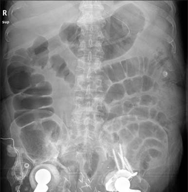
Fig. 1
Simple abdominal X-ray of consultation day. The film reveals dilatation of colon.
emj-37-52-g001.jpg
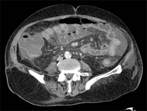
Fig. 2
Computed tomography of abdomen-pelvis. It shows edematous wall thickening with layered appearance from transverse colon to rectum with small ascites in pericolic area.
emj-37-52-g002.jpg
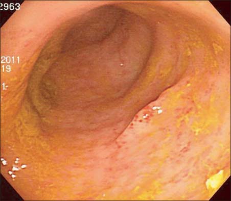
Fig. 3
Sigmoidoscopic finding. Sigmoidoscopy shows that normal colonic vasculature is disappeared and multiple erythematous erosions and atrophic changes are noted in colonic mucosa.
emj-37-52-g003.jpg
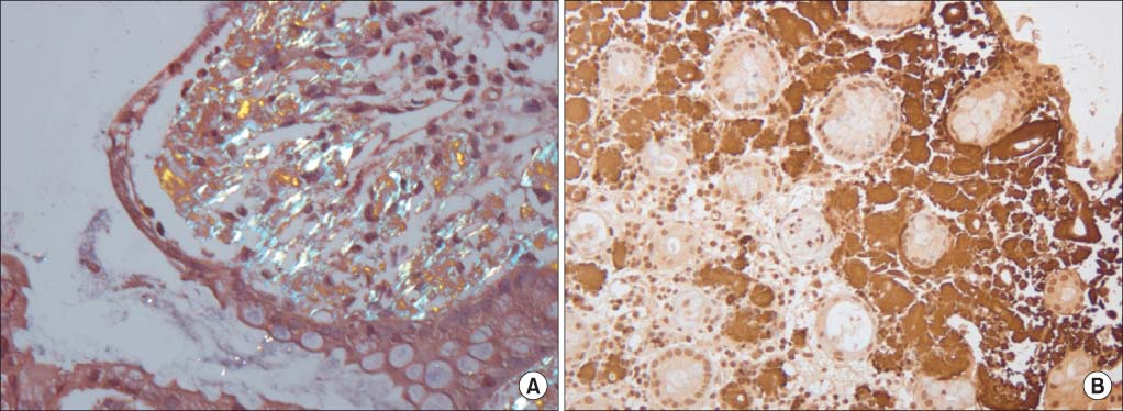
Fig. 4
Histopathologic fingings. (A) Congo-red stain reveals apple green birefringence in colonic stroma, compatible with amyloidosis (×100). (B) Immunohistochemical stain shows that amyloid A is strongly positive (×40).
emj-37-52-g004.jpg

Figure & Data

References

    Citations

    Citations to this article as recorded by  

      Download Citation

      Download a citation file in RIS format that can be imported by all major citation management software, including EndNote, ProCite, RefWorks, and Reference Manager.

      Format:

      Include:

      Toxic Megacolon Associated with Secondary Amyloidosis: An Unusual Complication of Clostridium difficile Colitis
      Ewha Med J. 2014;37(1):52-55.   Published online March 25, 2014
      Download Citation
      Download a citation file in RIS format that can be imported by all major citation management software, including EndNote, ProCite, RefWorks, and Reference Manager.

      Format:
      • RIS — For EndNote, ProCite, RefWorks, and most other reference management software
      • BibTeX — For JabRef, BibDesk, and other BibTeX-specific software
      Include:
      • Citation for the content below
      Toxic Megacolon Associated with Secondary Amyloidosis: An Unusual Complication of Clostridium difficile Colitis
      Ewha Med J. 2014;37(1):52-55.   Published online March 25, 2014
      Close

      Figure

      • 0
      • 1
      • 2
      • 3
      Toxic Megacolon Associated with Secondary Amyloidosis: An Unusual Complication of Clostridium difficile Colitis
      Image Image Image Image
      Fig. 1 Simple abdominal X-ray of consultation day. The film reveals dilatation of colon.
      Fig. 2 Computed tomography of abdomen-pelvis. It shows edematous wall thickening with layered appearance from transverse colon to rectum with small ascites in pericolic area.
      Fig. 3 Sigmoidoscopic finding. Sigmoidoscopy shows that normal colonic vasculature is disappeared and multiple erythematous erosions and atrophic changes are noted in colonic mucosa.
      Fig. 4 Histopathologic fingings. (A) Congo-red stain reveals apple green birefringence in colonic stroma, compatible with amyloidosis (×100). (B) Immunohistochemical stain shows that amyloid A is strongly positive (×40).
      Toxic Megacolon Associated with Secondary Amyloidosis: An Unusual Complication of Clostridium difficile Colitis
      TOP