• Contact us
  • E-Submission
ABOUT
BROWSE ARTICLES
JOURNAL POLICIES
FOR CONTRIBUTORS

Articles

Case Report

Adrenocortical Insufficiency due to Spontaneous Bilateral Adrenal Hemorrhage Presented as Acute Abdominal Pain

The Ewha Medical Journal 2014;37(Suppl):S10-S14. Published online: December 24, 2014

Department of Internal Medicine, Konyang University College of Medicine, Daejeon, Korea.

Corresponding author: Dong-Mee Lim. Department of Internal Medicine, Konyang University College of Medicine, 158 Gwanjeodong-ro, Seo-gu, Daejeon 302-718, Korea. Tel: 82-42-600-9169, Fax: 82-42-600-9090, mdldm@hanmail.net
• Received: May 12, 2014   • Accepted: August 6, 2014

Copyright © 2014, The Ewha Medical Journal

This is an Open Access article distributed under the terms of the Creative Commons Attribution Non-Commercial License (http://creativecommons.org/licenses/by-nc/3.0/) which permits unrestricted non-commercial use, distribution, and reproduction in any medium, provided the original work is properly cited.
  • 56 Views
  • 0 Download
  • 1 Crossref
prev next
  • Adrenocortical insufficiency is the clinical manifestation of deficient production or action of glucocorticoids. It is a life-threatening disorder that can result from primary adrenal failure or secondary adrenal failure due to impairment of the hypothalamic-pituitary axis. Primary adrenocortical insufficiency can be caused by autoimmune adrenalitis, infection (especially, tuberculosis), metastatic cancer, lymphoma, adrenal hemorrhage, infarction or drugs. Among these, adrenal hemorrhage may be caused by anticoagulant drug or heparin therapy, thromboembolic disease, hypercoagulable states such as antiphospholipid syndrome, physical trauma, postoperative state, sepsis and severe stress from any cause. However, even fewer reports exist of adrenocortical insufficiency due to spontaneous bilateral adrenal hemorrhage. We report a rare case of acute adrenocortical insufficiency due to spontaneous bilateral adrenal hemorrhage presenting as acute abdominal pain.
  • 1. Addison T. On the constitutional and local effects of disease of the supra-renal capsules; London: Samuel Highley; 1855.
  • 2. Charmandari E, Nicolaides NC, Chrousos GP. Adrenal insufficiency. Lancet 2014;383:2152-2167.
  • 3. Arlt W, Allolio B. Adrenal insufficiency. Lancet 2003;361:1881-1893.
  • 4. Rosenberger LH, Smith PW, Sawyer RG, Hanks JB, Adams RB, Hedrick TL. Bilateral adrenal hemorrhage: the unrecognized cause of hemodynamic collapse associated with heparin-induced thrombocytopenia. Crit Care Med 2011;39:833-838.
  • 5. Espinosa G, Santos E, Cervera R, Piette JC, de la Red G, Gil V, Font J, et al. Adrenal involvement in the antiphospholipid syndrome: clinical and immunologic characteristics of 86 patients. Medicine 2003;82:106-118.
  • 6. Rao RH, Vagnucci AH, Amico JA. Bilateral massive adrenal hemorrhage: early recognition and treatment. Ann Intern Med 1989;110:227-235.
  • 7. Feuerstein B, Streeten DH. Recovery of adrenal function after failure resulting from traumatic bilateral adrenal hemorrhages. Ann Intern Med 1991;115:785-786.
  • 8. Stewart PM, Krone NP. The adrenal cortex. Melmed S, Polonsky KS, Larsen PR, Kronenberg HM, editors. Williams textbook of endocrinology; 12th ed. Philadelphia, PA: Saunders Elsevier; 2011 479544.
  • 9. Xarli VP, Steele AA, Davis PJ, Buescher ES, Rios CN, Garcia-Bunuel R. Adrenal hemorrhage in the adult. Medicine 1978;57:211-221.
  • 10. Kovacs KA, Lam YM, Pater JL. Bilateral massive adrenal hemorrhage: assessment of putative risk factors by the case-control method. Medicine 2001;80:45-53.
  • 11. Swyer GI. Addison's disease. Br Med J 1979;2:25-26.
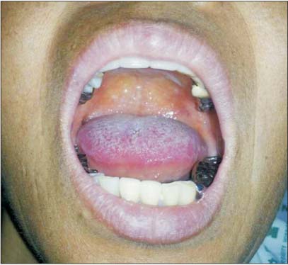
Fig. 1
Patient's tongue shows no hyperpigmentation.
emj-37-S10-g001.jpg
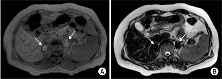
Fig. 2
Contrast-enhance abdominal computed tomography (CT). CT scan shows low-attenuation masses in both adrenal glands (arrow).
emj-37-S10-g002.jpg
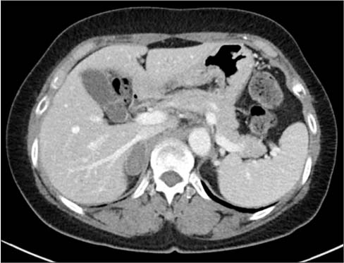
Fig. 3
Abdominal magnetic resonance imaging. Both adrenal masses show high signal intensity (arrow) on T1-weighted image (A) and low signal intensity (arrow) on T2-weighted image (B).
emj-37-S10-g003.jpg
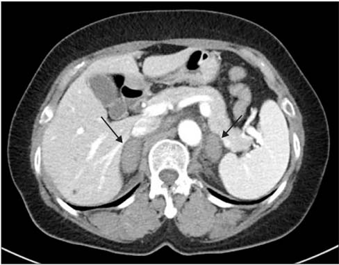
Fig. 4
Follow-up contrast-enhanced abdominal computed tomography (CT) after 2 weeks. CT scan shows decreased sizes of both adrenal hematomas. Also noted is rim-enhancement of right adrenal hematoma.
emj-37-S10-g004.jpg

Figure & Data

References

    Citations

    Citations to this article as recorded by  
    • A Case Report of Adrenal Insufficiency Treated with Korean Medicine
      Young-ji Kim, Jung-yeon Kwon, Ho-yeon Go, Kyung-hwan Kong
      The Journal of Internal Korean Medicine.2017; 38(5): 583.     CrossRef

    Download Citation

    Download a citation file in RIS format that can be imported by all major citation management software, including EndNote, ProCite, RefWorks, and Reference Manager.

    Format:

    Include:

    Adrenocortical Insufficiency due to Spontaneous Bilateral Adrenal Hemorrhage Presented as Acute Abdominal Pain
    Ewha Med J. 2014;37(Suppl):S10-S14.   Published online December 24, 2014
    Download Citation
    Download a citation file in RIS format that can be imported by all major citation management software, including EndNote, ProCite, RefWorks, and Reference Manager.

    Format:
    • RIS — For EndNote, ProCite, RefWorks, and most other reference management software
    • BibTeX — For JabRef, BibDesk, and other BibTeX-specific software
    Include:
    • Citation for the content below
    Adrenocortical Insufficiency due to Spontaneous Bilateral Adrenal Hemorrhage Presented as Acute Abdominal Pain
    Ewha Med J. 2014;37(Suppl):S10-S14.   Published online December 24, 2014
    Close

    Figure

    • 0
    • 1
    • 2
    • 3
    Adrenocortical Insufficiency due to Spontaneous Bilateral Adrenal Hemorrhage Presented as Acute Abdominal Pain
    Image Image Image Image
    Fig. 1 Patient's tongue shows no hyperpigmentation.
    Fig. 2 Contrast-enhance abdominal computed tomography (CT). CT scan shows low-attenuation masses in both adrenal glands (arrow).
    Fig. 3 Abdominal magnetic resonance imaging. Both adrenal masses show high signal intensity (arrow) on T1-weighted image (A) and low signal intensity (arrow) on T2-weighted image (B).
    Fig. 4 Follow-up contrast-enhanced abdominal computed tomography (CT) after 2 weeks. CT scan shows decreased sizes of both adrenal hematomas. Also noted is rim-enhancement of right adrenal hematoma.
    Adrenocortical Insufficiency due to Spontaneous Bilateral Adrenal Hemorrhage Presented as Acute Abdominal Pain
    TOP