• Contact us
  • E-Submission
ABOUT
BROWSE ARTICLES
JOURNAL POLICIES
FOR CONTRIBUTORS

Articles

Case Report

Actinomycosis Presented as Acute Appendicitis

The Ewha Medical Journal 2014;37(Suppl):S15-S18. Published online: December 24, 2014

Department of Internal Medicine, Ewha Womans University School of Medicine, Seoul, Korea.

1Department of Pathology, Ewha Womans University School of Medicine, Seoul, Korea.

Corresponding author: Hee Jung Choi. Department of Internal Medicine, Ewha Womans University School of Medicine, 1071 Anyancheon-ro, Yangcheon-gu, Seoul 158-710, Korea. Tel: 82-2-2650-5272, Fax: 82-2-2655-2076, heechoi@ewha.ac.kr
• Received: May 29, 2014   • Accepted: July 23, 2014

Copyright © 2014, The Ewha Medical Journal

This is an Open Access article distributed under the terms of the Creative Commons Attribution Non-Commercial License (http://creativecommons.org/licenses/by-nc/3.0/) which permits unrestricted non-commercial use, distribution, and reproduction in any medium, provided the original work is properly cited.
  • 37 Views
  • 0 Download
prev next
  • Actinomycosis causes a chronic suppurative, granulomatous disease which is characterized by extensive abscess formation, and sulfur granule formation. Actinomycosis may present different clinical forms: cervicofacial, thoracic, abdominal and cerebral actinomycosis. The diagnosis can only be made after surgery. In general, patients with abdominal actinomycosis have undergone abdominal surgery. We report four cases of primary appendiceal actinomycosis presenting as acute appendicitis without history of abdomen surgery.
  • 1. Lunca S, Bouras G, Romedea NS, Pertea M. Abdominal wall actinomycosis associated with prolonged use of an intrauterine device: a case report and review of the literature. Int Surg 2005;90:236-240.
  • 2. Karagulle E, Turan H, Turk E, Kiyici H, Yildirim E, Moray G. Abdominal actinomycosis mimicking acute appendicitis. Can J Surg 2008;51:E109-E110.
  • 3. Hsu JT, Lo HC, Jan YY, Chen HM. Actinomycosis mimicking recurrent carcinoma after Whipple's operation. World J Gastroenterol 2005;11:1722-1724.
  • 4. Sudhakar SS, Ross JJ. Short-term treatment of actinomycosis: two cases and a review. Clin Infect Dis 2004;38:444-447.
  • 5. Yeung VH, Wong QH, Chao NS, Leung MW, Kwok WK. Thoracic actinomycosis in an adolescent mimicking chest wall tumor or pulmonary tuberculosis. Pediatr Surg Int 2008;24:751-754.
  • 6. Vyas JM, Kasmar A, Chang HR, Holden J, Hohmann E. Abdominal abscesses due to actinomycosis after laparoscopic cholecystectomy: case reports and review. Clin Infect Dis 2007;44:e1-e4.
  • 7. Lee SY, Kwon HJ, Cho JH, Oh JY, Nam KJ, Lee JH, et al. Actinomycosis of the appendix mimicking appendiceal tumor: a case report. World J Gastroenterol 2010;16:395-397.
  • 8. Abela J, Sciberras J, Meilak M, Felice AG, Degaetano J. Omental actinomycosis presenting with right lower quadrant abdominal pain. J Clin Pathol 2004;57:671.
  • 9. Mandell GL, Bennett JE, Dolin R. Mandell, Douglas, and Bennett's principles and practices of infectious diseases; 7th ed. Cambridge: Churchill Livingstone; 2009.
  • 10. Sevilla CF, Villalba FF, Domingo DPC, Laforga CJ, de La Morena VE. Abdominal actinomycosis simulating Crohn's disease. Gastroenterol Hepatol 2001;24:300-302.
  • 11. Huang CJ, Huang TJ, Hsieh JS. Pseudo-colonic carcinoma caused by abdominal actinomycosis: report of two cases. Int J Colorectal Dis 2004;19:283-286.
  • 12. Garcia Garcia JC, Nunez Fernandez MJ, Cerqueiro Gonzalez JM, Garcia Martin C, Rodriguez Garcia JC, Anibarro Garcia L, et al. Primary actinomycosis of the abdominal wall: description of 2 cases and review of the literature. An Med Interna 2001;18:80-83.
  • 13. Ferrari TC, Couto CA, Murta-Oliveira C, Conceicao SA, Silva RG. Actinomycosis of the colon: a rare form of presentation. Scand J Gastroenterol 2000;35:108-109.
  • 14. Liu V, Val S, Kang K, Velcek F. Case report: actinomycosis of the appendix--an unusual cause of acute appendicitis in children. J Pediatr Surg 2010;45:2050-2052.
  • 15. Choi MM, Baek JH, Lee JN, Park S, Lee WS. Clinical features of abdominopelvic actinomycosis: report of twenty cases and literature review. Yonsei Med J 2009;50:555-559.
Fig. 1
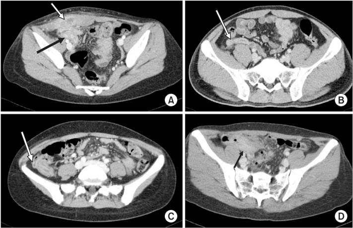
Computed tomography (CT) of abdomen-pelvis. (A) It shows thickening of appendiceal wall with periappendiceal soft tissue (black arrow) and right rectus muscle infiltration (white arrow). (B, C) CT scan shows appendiceal wall thickening with enhancement (arrow). (D) It shows acute appendicitis with perforation and abscess formation (arrow).
emj-37-S15-g001.jpg
Fig. 2
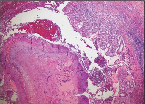
Histopathologic findings. A large actinomycotic granule was embedded in acute suppurative inflammatory exudates in the appendiceal lumen. Food material presented at the center of the granule as a nidus (H&E, ×40).
emj-37-S15-g002.jpg
Fig. 3
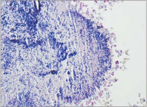
Histopathologic fingings. Tangled and irregular aggregates of delicate filamentous gram-positive bacilli are embedded within the matrix of the granule (Gram stain, ×400).
emj-37-S15-g003.jpg

Figure & Data

Fig. 1
Computed tomography (CT) of abdomen-pelvis. (A) It shows thickening of appendiceal wall with periappendiceal soft tissue (black arrow) and right rectus muscle infiltration (white arrow). (B, C) CT scan shows appendiceal wall thickening with enhancement (arrow). (D) It shows acute appendicitis with perforation and abscess formation (arrow).
emj-37-S15-g001.jpg
Fig. 2
Histopathologic findings. A large actinomycotic granule was embedded in acute suppurative inflammatory exudates in the appendiceal lumen. Food material presented at the center of the granule as a nidus (H&E, ×40).
emj-37-S15-g002.jpg
Fig. 3
Histopathologic fingings. Tangled and irregular aggregates of delicate filamentous gram-positive bacilli are embedded within the matrix of the granule (Gram stain, ×400).
emj-37-S15-g003.jpg

References

  • 1. Lunca S, Bouras G, Romedea NS, Pertea M. Abdominal wall actinomycosis associated with prolonged use of an intrauterine device: a case report and review of the literature. Int Surg 2005;90:236-240.
  • 2. Karagulle E, Turan H, Turk E, Kiyici H, Yildirim E, Moray G. Abdominal actinomycosis mimicking acute appendicitis. Can J Surg 2008;51:E109-E110.
  • 3. Hsu JT, Lo HC, Jan YY, Chen HM. Actinomycosis mimicking recurrent carcinoma after Whipple's operation. World J Gastroenterol 2005;11:1722-1724.
  • 4. Sudhakar SS, Ross JJ. Short-term treatment of actinomycosis: two cases and a review. Clin Infect Dis 2004;38:444-447.
  • 5. Yeung VH, Wong QH, Chao NS, Leung MW, Kwok WK. Thoracic actinomycosis in an adolescent mimicking chest wall tumor or pulmonary tuberculosis. Pediatr Surg Int 2008;24:751-754.
  • 6. Vyas JM, Kasmar A, Chang HR, Holden J, Hohmann E. Abdominal abscesses due to actinomycosis after laparoscopic cholecystectomy: case reports and review. Clin Infect Dis 2007;44:e1-e4.
  • 7. Lee SY, Kwon HJ, Cho JH, Oh JY, Nam KJ, Lee JH, et al. Actinomycosis of the appendix mimicking appendiceal tumor: a case report. World J Gastroenterol 2010;16:395-397.
  • 8. Abela J, Sciberras J, Meilak M, Felice AG, Degaetano J. Omental actinomycosis presenting with right lower quadrant abdominal pain. J Clin Pathol 2004;57:671.
  • 9. Mandell GL, Bennett JE, Dolin R. Mandell, Douglas, and Bennett's principles and practices of infectious diseases; 7th ed. Cambridge: Churchill Livingstone; 2009.
  • 10. Sevilla CF, Villalba FF, Domingo DPC, Laforga CJ, de La Morena VE. Abdominal actinomycosis simulating Crohn's disease. Gastroenterol Hepatol 2001;24:300-302.
  • 11. Huang CJ, Huang TJ, Hsieh JS. Pseudo-colonic carcinoma caused by abdominal actinomycosis: report of two cases. Int J Colorectal Dis 2004;19:283-286.
  • 12. Garcia Garcia JC, Nunez Fernandez MJ, Cerqueiro Gonzalez JM, Garcia Martin C, Rodriguez Garcia JC, Anibarro Garcia L, et al. Primary actinomycosis of the abdominal wall: description of 2 cases and review of the literature. An Med Interna 2001;18:80-83.
  • 13. Ferrari TC, Couto CA, Murta-Oliveira C, Conceicao SA, Silva RG. Actinomycosis of the colon: a rare form of presentation. Scand J Gastroenterol 2000;35:108-109.
  • 14. Liu V, Val S, Kang K, Velcek F. Case report: actinomycosis of the appendix--an unusual cause of acute appendicitis in children. J Pediatr Surg 2010;45:2050-2052.
  • 15. Choi MM, Baek JH, Lee JN, Park S, Lee WS. Clinical features of abdominopelvic actinomycosis: report of twenty cases and literature review. Yonsei Med J 2009;50:555-559.

Citations

Citations to this article as recorded by  
    CanvasJS.com

    Download Citation

    Download a citation file in RIS format that can be imported by all major citation management software, including EndNote, ProCite, RefWorks, and Reference Manager.

    Format:

    Include:

    Actinomycosis Presented as Acute Appendicitis
    Ewha Med J. 2014;37(Suppl):S15-S18.   Published online December 24, 2014
    Download Citation
    Download a citation file in RIS format that can be imported by all major citation management software, including EndNote, ProCite, RefWorks, and Reference Manager.

    Format:
    • RIS — For EndNote, ProCite, RefWorks, and most other reference management software
    • BibTeX — For JabRef, BibDesk, and other BibTeX-specific software
    Include:
    • Citation for the content below
    Actinomycosis Presented as Acute Appendicitis
    Ewha Med J. 2014;37(Suppl):S15-S18.   Published online December 24, 2014
    Close

    Figure

    • 2
    • 0
    • 1
    • 2
    • 0
    Actinomycosis Presented as Acute Appendicitis
    Image Image Image
    Fig. 1 Computed tomography (CT) of abdomen-pelvis. (A) It shows thickening of appendiceal wall with periappendiceal soft tissue (black arrow) and right rectus muscle infiltration (white arrow). (B, C) CT scan shows appendiceal wall thickening with enhancement (arrow). (D) It shows acute appendicitis with perforation and abscess formation (arrow).
    Fig. 2 Histopathologic findings. A large actinomycotic granule was embedded in acute suppurative inflammatory exudates in the appendiceal lumen. Food material presented at the center of the granule as a nidus (H&E, ×40).
    Fig. 3 Histopathologic fingings. Tangled and irregular aggregates of delicate filamentous gram-positive bacilli are embedded within the matrix of the granule (Gram stain, ×400).
    Actinomycosis Presented as Acute Appendicitis
    TOP Mpgyi