• Contact us
  • E-Submission
ABOUT
BROWSE ARTICLES
JOURNAL POLICIES
FOR CONTRIBUTORS

Articles

Page Path

Case Report

Ovarian Metastasis from Non-Small Cell Lung Cancer Responding to Erlotinib

The Ewha Medical Journal 2015;38(1):46-49. Published online: March 26, 2015

Department of Internal Medicine, Konyang University College of Medicine, Daejeon, Korea.

Corresponding author: Do Yeun Cho. Department of Internal Medicine, Konyang University College of Medicine, 158 Gwanjeodong-ro, Seo-gu, Daejeon 302-718, Korea. Tel: 82-42-600-9102, Fax: 82-42-600-9092, dycho@kyuh.ac.kr
• Received: August 15, 2014   • Accepted: September 26, 2014

Copyright © 2015, The Ewha Medical Journal

This is an Open Access article distributed under the terms of the Creative Commons Attribution Non-Commercial License (http://creativecommons.org/licenses/by-nc/3.0/) which permits unrestricted non-commercial use, distribution, and reproduction in any medium, provided the original work is properly cited.
  • 27 Views
  • 0 Download
prev next
  • Ovarian cancer is generally primary cancer and less frequently originates from metastasis from non-gynecological cancer. Ovarian metastasis from lung cancer represents only 2~4% of all ovarian metastatic cancers. We report a case of ovarian metastasis of non-small cell lung cancer with epidermal growth factor receptor mutation. The patient underwent cytoreductive surgery for the ovarian mass and erlotinib therapy for the metastatic lung cancer. Erlotinib therapy markedly decreased the size of lung mass.
  • 1. Hart W. Diagnostic challenge of secondary (metastatic) ovarian tumors simulating primary endometrioid and mucinous neoplasms. Pathol Int 2005;55:231-243.
  • 2. Horn L, Pao W, Johnson D. Neoplasm of the Lung. Longo D, Fauci A, Kasper D, Hauser S, Jameson J, Loscalzo J, editors. Harrison's principles of internal medicine; 18th ed. New York: McGraw Hill; 2012 745750.
  • 3. Schiller J, Harrington D, Belani C, Langer C, Sandler A, Krook J, et al. Comparison of four chemotherapy regimens for advanced non-small-cell lung cancer. N Engl J Med 2002;346:92-98.
  • 4. Lynch T, Bell D, Sordella R, Gurubhagavatula S, Okimoto R, Brannigan B, et al. Activating mutations in the epidermal growth factor receptor underlying responsiveness of non-small-cell lung cancer to gefetinib. N Engl J Med 2004;350:2129-2139.
  • 5. Fader A, Rose P. Role of surgery in ovarian carcinoma. J Clin Oncol 2007;25:2873-2883.
  • 6. Jung YE, Lee JW, Kim BG, Bae DS. Ovarian metastasis from pulmonary adenocarcinoma. Obstet Gynecol Sci 2013;56:341-344.
  • 7. Varadhachary G, Abbruzzese J. Carcinoma of Unknown Primary. Longo D, Fauci A, Kasper D, Hauser S, Jameson J, Loscalzo J, editors. Harrison's principles of internal medicine; 18th ed. New York: McGraw Hill; 2012 822823.
  • 8. Pfister D, Johnson D, Azzoli C, Sause W, Smith T, Baker S, et al. American society of clinical oncology treatment of unresectable non-small-cell lung cancer guideline: update 2003. J Clin Oncol 2004;22:330-353.
  • 9. Hirsch F, Varella-Garcia M, Bunn P Jr, Franklin W, Dziadziuszko R, Thatcher N, et al. Molecular predictors of outcome with gefetinib in a phase III placebo-controlled study in advanced nonsmall-cell lung cancer. J Clin Oncol 2006;24:5034-5042.
  • 10. Sequist L, Bell D, Lynch T, Haber D. Molecular predictors of response to epidermal growth factor receptor antagonists in nonsmall-cell lung cancer. J Clin Oncol 2007;25:587-595.
Fig. 1

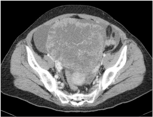
Abdomen and pelvis computed tomography. A 14.7×13.4 cm sized huge tumor of right ovary and peritoneal carcinomatosis are noted.

emj-38-46-g001.jpg
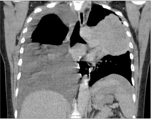
Fig. 2

Chest computed tomography. It reveals a 10×6 cm sized huge mass in left upper lung and massive right sided pleural effusion.

emj-38-46-g002.jpg
Fig. 3

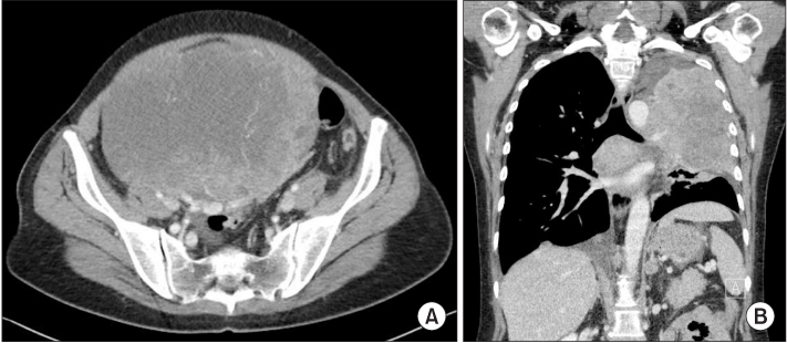
Images taken after 2 cycles of gemcitabine with carboplatin chemotherapy. (A) Abdomen and pelvis computed tomography (CT) reveals increasing the size of left ovarian mass (20×14.5 cm). (B) Chest CT reveals increasing the size of left upper lung mass (12×8.2 cm).

emj-38-46-g003.jpg
Fig. 4

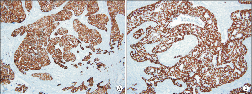
Immunohistochemical staining of the left ovary. (A) Cytokeratin-7 staining reveals strong reactivity (×100). (B) Thyroid transcription factor-1 staining reveals strong reactivity (×100).

emj-38-46-g004.jpg
Fig. 5

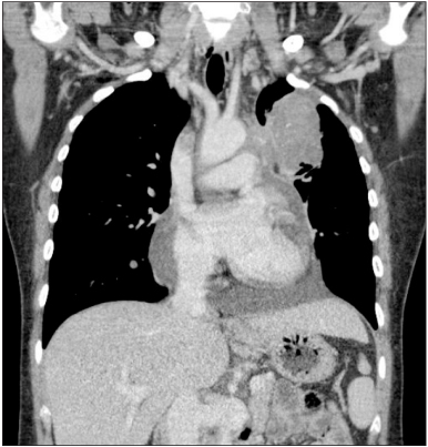
Chest CT after erlotinib chemotherapy. It reveals a marked decrease in the size of the left upper lung mass (4.2×3.7 cm).

emj-38-46-g005.jpg

Figure & Data

References

    Citations

    Citations to this article as recorded by  

      Download Citation

      Download a citation file in RIS format that can be imported by all major citation management software, including EndNote, ProCite, RefWorks, and Reference Manager.

      Format:

      Include:

      Ovarian Metastasis from Non-Small Cell Lung Cancer Responding to Erlotinib
      Ewha Med J. 2015;38(1):46-49.   Published online March 26, 2015
      Download Citation
      Download a citation file in RIS format that can be imported by all major citation management software, including EndNote, ProCite, RefWorks, and Reference Manager.

      Format:
      • RIS — For EndNote, ProCite, RefWorks, and most other reference management software
      • BibTeX — For JabRef, BibDesk, and other BibTeX-specific software
      Include:
      • Citation for the content below
      Ovarian Metastasis from Non-Small Cell Lung Cancer Responding to Erlotinib
      Ewha Med J. 2015;38(1):46-49.   Published online March 26, 2015
      Close

      Figure

      • 0
      • 1
      • 2
      • 3
      • 4
      Ovarian Metastasis from Non-Small Cell Lung Cancer Responding to Erlotinib
      Image Image Image Image Image
      Fig. 1 Abdomen and pelvis computed tomography. A 14.7×13.4 cm sized huge tumor of right ovary and peritoneal carcinomatosis are noted.
      Fig. 2 Chest computed tomography. It reveals a 10×6 cm sized huge mass in left upper lung and massive right sided pleural effusion.
      Fig. 3 Images taken after 2 cycles of gemcitabine with carboplatin chemotherapy. (A) Abdomen and pelvis computed tomography (CT) reveals increasing the size of left ovarian mass (20×14.5 cm). (B) Chest CT reveals increasing the size of left upper lung mass (12×8.2 cm).
      Fig. 4 Immunohistochemical staining of the left ovary. (A) Cytokeratin-7 staining reveals strong reactivity (×100). (B) Thyroid transcription factor-1 staining reveals strong reactivity (×100).
      Fig. 5 Chest CT after erlotinib chemotherapy. It reveals a marked decrease in the size of the left upper lung mass (4.2×3.7 cm).
      Ovarian Metastasis from Non-Small Cell Lung Cancer Responding to Erlotinib
      TOP