• Contact us
  • E-Submission
ABOUT
BROWSE ARTICLES
JOURNAL POLICIES
FOR CONTRIBUTORS

Articles

Page Path

Case Report

Rectus Abdominis Muscle Tuberculosis Involving Lung and Endometrium

The Ewha Medical Journal 2015;38(2):76-79. Published online: July 29, 2015

Department of Internal Medicine, Ewha Womans University School of Medicine, Seoul, Korea.

1Department of Radiology, Ewha Womans University School of Medicine, Seoul, Korea.

2Department of Pathology, Ewha Womans University School of Medicine, Seoul, Korea.

3Department of Obstetrics and Gynecology, Ewha Womans University School of Medicine, Seoul, Korea.

Corresponding author: Eun Mi Chun. Division of Pulmonology, Department Internal Medicine, Ewha Womans University School of Medicine, 1071 Anyangcheon-ro, Yangcheon-gu, Seoul 158-710, Korea. Tel: 82-2-2650-5131, Fax: 82-2-2650-2559, cem@ewha.ac.kr
• Received: April 3, 2015   • Accepted: June 3, 2015

Copyright © 2015, The Ewha Medical Journal

This is an Open Access article distributed under the terms of the Creative Commons Attribution Non-Commercial License (http://creativecommons.org/licenses/by-nc/3.0/) which permits unrestricted non-commercial use, distribution, and reproduction in any medium, provided the original work is properly cited.
  • 41 Views
  • 0 Download
prev next
  • Extrapulmonary tuberculosis usually involves the pleura, central nervous system, lymphatic system, genitourinary system and bone with joint. There are few reports about pulmonary tuberculosis involving the endometrium and muscle. A 32-year-old woman who had intact immunity visited hospital due to menorrhagia, and was diagnosed with endometrial tuberculosis. The patient also had a painless abdominal mass about for 1 year before she came to the hospital. She was diagnosed with rectus abdominis muscle tuberculosis, endometrial tuberculosis, and pulmonary tuberculosis at the same time. We report a case of endometrial tuberculosis accompanied with rectus abdominis muscle and pulmonary tuberculosis in a non-immunosuppressed person.
  • 1. Sharma SK, Mohan A. Extrapulmonary tuberculosis. Indian J Med Res 2004;120:316-353.
  • 2. Wang WY, Lin FC, Tsao TY, Lu JJ. Tuberculous myositis: an unusual presentation of extrapulmonary tuberculosis. J Microbiol Immunol Infect 2007;40:79-82.
  • 3. Parikh FR, Nadkarni SG, Kamat SA, Naik N, Soonawala SB, Parikh RM. Genital tuberculosis: a major pelvic factor causing infertility in Indian women. Fertil Steril 1997;67:497-500.
  • 4. Weir MR, Thornton GF. Extrapulmonary tuberculosis: experience of a community hospital and review of the literature. Am J Med 1985;79:467-478.
  • 5. Nuwal P, Dixit R. Tuberculosis of rectus abdominis muscle. Indian J Chest Dis Allied Sci 2007;49:239-240.
  • 6. Sabat D, Kumar V. Primary tuberculous abscess of rectus femoris muscle: a case report. J Infect Dev Ctries 2009;3:476-478.
  • 7. Nuwal P, Dixit R. Tuberculosis of rectus abdominis muscle. Indian J Chest Dis Allied Sci 2007;49:239-240.
  • 8. World Health Organization.Global tuberculosis control: surveillance, planning, financing. WHO report 2007; Geneva: World Health Organization; 2007.
  • 9. Chavhan GB, Hira P, Rathod K, Zacharia TT, Chawla A, Badhe P, et al. Female genital tuberculosis: hysterosalpingographic appearances. Br J Radiol 2004;77:164-169.
  • 10. Nabag WO, Nur Hassan A, Sayed DM, El Sheikh MA. endometrial tuberculosis and secondary amenorrhea: a report of three cases in Sudan. J Minim Invasive Surg Sci 2012;1:30-33.
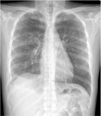
Fig. 1

Chest X-ray on admission. It shows a high probability of active tuberculosis in right lung with tuberculous empyema in right lower hemithorax.

emj-38-76-g001.jpg
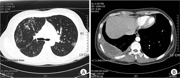
Fig. 2

Chest computed tomography scan. It shows multifocal centrilobular nodule, 'tree in bud appearance' with consolidation on both upper lobes (A). Chest computed tomography scan shows tuberculous empyema in right lower hemithorax (B).

emj-38-76-g002.jpg
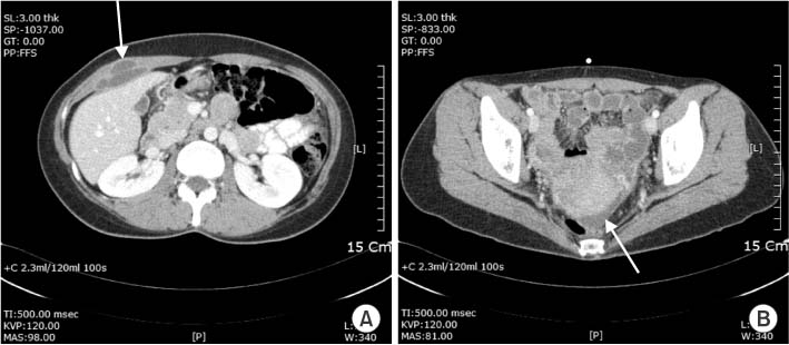
Fig. 3

Abdomino-pelvic computed tomography (APCT) scan on admission. It shows a well-defined cystic mass with wall formation in right rectus abdominis muscle (5×2 cm) (A). APCT scan shows irregular thickening and enhancement of pelvic parietal peritoneum (B).

emj-38-76-g003.jpg
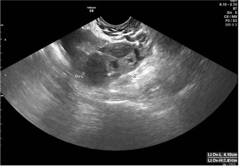
Fig. 4

Finding of transvaginal ultrasonography. It shows left ovarian complex cyst, which cannot completely exclude tuberculoma (Ov-L: ovary length; Ov-H: ovary height).

emj-38-76-g004.jpg
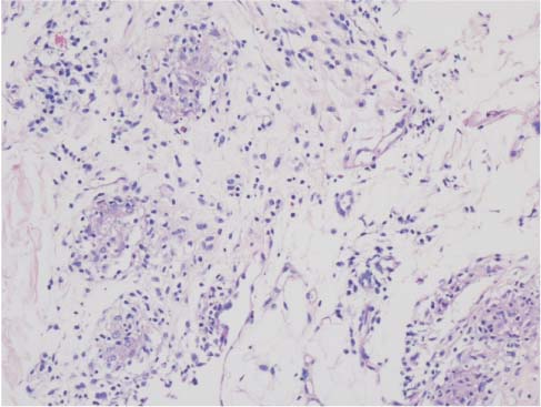
Fig. 5

Histopathologic finding of rectus abdominis muscle. It shows chronic granulomatous inflammation, suggestive of tuberculosis (H&E, ×200).

emj-38-76-g005.jpg

Figure & Data

References

    Citations

    Citations to this article as recorded by  

      Download Citation

      Download a citation file in RIS format that can be imported by all major citation management software, including EndNote, ProCite, RefWorks, and Reference Manager.

      Format:

      Include:

      Rectus Abdominis Muscle Tuberculosis Involving Lung and Endometrium
      Ewha Med J. 2015;38(2):76-79.   Published online July 29, 2015
      Download Citation
      Download a citation file in RIS format that can be imported by all major citation management software, including EndNote, ProCite, RefWorks, and Reference Manager.

      Format:
      • RIS — For EndNote, ProCite, RefWorks, and most other reference management software
      • BibTeX — For JabRef, BibDesk, and other BibTeX-specific software
      Include:
      • Citation for the content below
      Rectus Abdominis Muscle Tuberculosis Involving Lung and Endometrium
      Ewha Med J. 2015;38(2):76-79.   Published online July 29, 2015
      Close

      Figure

      • 0
      • 1
      • 2
      • 3
      • 4
      Rectus Abdominis Muscle Tuberculosis Involving Lung and Endometrium
      Image Image Image Image Image
      Fig. 1 Chest X-ray on admission. It shows a high probability of active tuberculosis in right lung with tuberculous empyema in right lower hemithorax.
      Fig. 2 Chest computed tomography scan. It shows multifocal centrilobular nodule, 'tree in bud appearance' with consolidation on both upper lobes (A). Chest computed tomography scan shows tuberculous empyema in right lower hemithorax (B).
      Fig. 3 Abdomino-pelvic computed tomography (APCT) scan on admission. It shows a well-defined cystic mass with wall formation in right rectus abdominis muscle (5×2 cm) (A). APCT scan shows irregular thickening and enhancement of pelvic parietal peritoneum (B).
      Fig. 4 Finding of transvaginal ultrasonography. It shows left ovarian complex cyst, which cannot completely exclude tuberculoma (Ov-L: ovary length; Ov-H: ovary height).
      Fig. 5 Histopathologic finding of rectus abdominis muscle. It shows chronic granulomatous inflammation, suggestive of tuberculosis (H&E, ×200).
      Rectus Abdominis Muscle Tuberculosis Involving Lung and Endometrium
      TOP