• Contact us
  • E-Submission
ABOUT
BROWSE ARTICLES
JOURNAL POLICIES
FOR CONTRIBUTORS

Articles

Page Path

Orginal Article

Characteristics for Ischemic Stroke in 18–30 Years Old Patients, Multicenter Stroke Registry Study

The Ewha Medical Journal 2017;40(3):128-135. Published online: July 28, 2017

Department of Neurology, Ewha Womans University School of Medicine, Seoul, Korea

1Departments of Neurology, Yonsei University College of Medicine, Seoul, Korea

2Department of Neurology, Hallym University College of Medicine, Sacred Heart Hospital, Hallym University College of Medicine, Anyang, Korea

33Department of Neurology, Dongtan Sacred Heart Hospital, Hallym University College of Medicine, Hwaseong, Korea

Corresponding author Suk Yun Kang Department of Neurology, Dongtan Sacred Heart Hospital, Hallym University College of Medicine, 7 Keunjaebong-gil, Hwaseong 18450, Korea Tel: 82-31-8086-2310, Fax: 82-31-8086-2317 E-mail: sukyunkang@hanmail.net
• Received: January 15, 2017   • Revised: April 20, 2017   • Accepted: April 26, 2017

Copyright © 2017 Ewha Womans University School of Medicine

This is an Open Access article distributed under the terms of the Creative Commons Attribution Non-Commercial License (http://creativecommons.org/licenses/by-nc/4.0/) which permits unrestricted non-commercial use, distribution, and reproduction in any medium, provided the original work is properly cited.
  • 43 Views
  • 0 Download
prev next
  • Objectives
    Although there have been several reports that described characteristics for young age stroke, information regarding very young age (18–30 years old) has been limited. We aimed to analyze demographic factors, stroke subtype, and 3-month outcome in acute ischemic stroke patient who have relatively very young age in multicenter stroke registry.
  • Methods
    We evaluated all 122 (7.1%) consecutive acute ischemic stroke (within 7 days after symptom onset) patients aged 18 to 30 from 17,144 patients who registered in multicenter prospective stroke registry, 1997 to 2012. Etiology was classified by Trial of Org 10172 in Acute Stroke Treatment criteria. Stroke severity was defined as National Institutes of Health Stroke Scale (NIHSS) and stroke outcome was defined by modified Rankin scale (mRS) at 3 months after index stroke.
  • Results
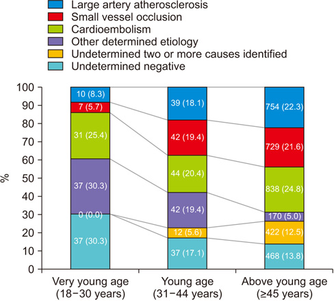
    The mean age of all included patients was 25.1±3.7 years and 76 patients (62.2%) were male. The median NIHSS at admission was 4. Considering stroke subtype, 37 patients (30.3%) had stroke of other determined etiology (SOD), 37 (30.3%) had undetermined negative evaluation (UN) and 31 (25.4%) had cardioembolism (CE) were frequently noted. After adjusting age, sex and variables which had P<0.1 in univariable analysis (NIHSS and stroke subtype), CE stroke subtype (odds ratio, 4.68; 95% confidence interval, 1.42–15.48; P=0.011) were significantly associated with poor functional outcome (mRS≥3).
  • Conclusion
    In very young age ischemic stroke patients, SOD and UN stroke subtype were most common and CE stroke subtype was independently associated with poor discharge outcome.
Fig. 1.
Stroke subtype comparison for very young age (18–30 years old) with young age (31–44 years old) or above young age (≥45 years old). Values are presented as number (%). The stroke of other determined etiology and undetermined negative evaluation stroke subtype were more frequently noted in 18–30 years old patients than those of 31–44 years old and ≥45 years old. The frequency of large artery atherosclerosis and small vessel occlusion subtypes was increased in elderly groups (31–44 years old and ≥45 years old groups; P=0.001).
emj-2017-40-3-128f1.jpg
Table 1.
Demographic and clinical data of included patients
Variable mRS<3 (n=95) mRS≥3 (n=27) Total (N=122) P-value
Male sex 61 (64.2) 15 (55.6) 76 (62.3) 0.413
Age (yr) 25.1±3.8 24.8±3.3 25.1±3.7 0.646
Risk factor
Hypertension 12 (12.6) 3 (11.1) 15 (12.3) 0.832
Diabetes mellitus 1 (1.1) 0 1 (0.8) 0.592
Dyslipidemia 8 (8.4) 0 8 (6.6) 0.119
Smoking 45 (47.4) 9 (33.3) 54 (44.3) 0.195
Regular alcohol intake 23 (24.2) 7 (25.9) 30 (24.6) 0.855
Recent alcohol intake 8 (8.4) 0 8 (6.6) 0.126
Atrial fibrillation 2 (2.1) 5 (18.5) 7 (5.7) 0.001
Occupation situation 0.140
Without routine job 13 (13.7) 7 (25.9) 20 (16.4)
White collar 37 (38.9) 4 (18.9) 41 (33.6)
Soldier 22 (23.2) 6 (22.2) 28 (23.0)
Student 20 (21.1) 9 (33.3) 29 (23.8)
Blue collar 3 (3.2) 1 (3.7) 4 (3.3)
Activity at stroke occurrence 0.183
Resting 68 (71.6) 23 (85.2) 91 (74.6)
Vigour activity 17 (17.9) 1 (3.7) 18 (14.8)
Wake-up stroke 10 (10.5) 3 (11.1) 13 (10.7)
Stroke classification 0.028
Large artery atherosclerosis 8 (8.4) 2 (7.5) 10 (8.3)
Small vessel occlusion 7 (7.4) 0 7 (5.7)
Cardioembolism 18 (18.9) 13 (48.1) 31 (25.4)
Other determined etiology 30 (31.6) 7 (25.9) 37 (30.3)
Undetermined negative 32 (33.7) 5 (18.5) 37 (30.3)
NIHSS 3 (1–5) 12 (9–14) 4 (2–8) 0.001

Values are presented as mean±standard deviation, number (%), or median (interquartile range).

mRS, modified Rankin scale at 3 months after index stroke; NIHSS, National Institute of Health Stroke Scale.

Table 2.
Causes for other determined etiology
Variable Causes for other determined etiology (n=37)
Arterial dissection 21 (56.8)
 Extracranial internal carotid artery 8 (21.6)
 Intracranial internal carotid artery* 1 (2.7)
 Extracranial vertebral artery 6 (16.2)
 Intracranial vertebral artery 3 (8.1)
 Middle cerebral artery 3 (8.1)
Vasculitis meningitis 4 (10.8)
Moyamoya diseases 4 (10.8)
Takayasu’s diseases 3 (8.1)
Systemic lupus erythematosus 3 (8.1)
Sneddon’s syndrome 1 (2.7)
Familial protein C, S deficiency 1 (2.7)

Values are presented as number (%).

*One patient had both right intracranial internal carotid artery and middle cerebral artery dissection.

Table 3.
Independent factors for poor functional outcome at 3 months (mRS≥3)
Variable Adjusted OR (95% confidence interval) P-value
Male sex 1.41 (0.56–3.54) 0.455
Age (yr) 0.96 (0.84–1.09) 0.564
NIHSS 2.94 (1.61–5.37) 0.001
Stroke subtype
 Large artery atherosclerosis 1.53 (0.23–9.91) 0.652
 Small vessel occlusion No case
 Cardioembolism 4.68 (1.42–15.48) 0.011
 Other determined etiology 1.37 (0.38–4.86) 0.626
 Undetermined negative Reference

mRS, modified Rankin scale; OR, odds ratio; NIHSS, National Institute of Health Stroke Scale.

  • 1. Kristensen B, Malm J, Carlberg B, Stegmayr B, Backman C, Fagerlund M, et al. Epidemiology and etiology of ischemic stroke in young adults aged 18 to 44 years in northern Sweden. Stroke 1997;28:1702-1709.
  • 2. Carolei A, Marini C, Ferranti E, Frontoni M, Prencipe M, Fieschi C. A prospective study of cerebral ischemia in the young: analysis of pathogenic determinants. The National Research Council Study Group. Stroke 1993;24:362-367.
  • 3. Siqueira Neto JI, Santos AC, Fabio SR, Sakamoto AC. Cerebral infarction in patients aged 15 to 40 years. Stroke 1996;27:2016-2019.
  • 4. Hong KS, Bang OY, Kang DW, Yu KH, Bae HJ, Lee JS, et al. Stroke statistics in Korea: part I. Epidemiology and risk factors: a report from the Korean Stroke Society and Clinical Research Center For Stroke. J Stroke 2013;15:2-20.
  • 5. Kwon SU, Kim JS, Lee JH, Lee MC. Ischemic stroke in Korean young adults. Acta Neurol Scand 2000;101:19-24.
  • 6. Tibæk M, Dehlendorff C, Jørgensen HS, Forchhammer HB, Johnsen SP, Kammersgaard LP. Increasing incidence of hospitalization for stroke and transient ischemic attack in young adults: a registry-based study. J Am Heart Assoc 2016;5:e003158.
  • 7. Song TJ, Suh SH, Min PK, Kim DJ, Kim BM, Heo JH, et al. The influence of anti-platelet resistance on the development of cerebral ischemic lesion after carotid artery stenting. Yonsei Med J 2013;54:288-294.
  • 8. Lee BI, Nam HS, Heo JH, Kim DI. Yonsei Stroke Team. Yonsei Stroke Registry: analysis of 1,000 patients with acute cerebral infarctions. Cerebrovasc Dis 2001;12:145-151.
  • 9. Song TJ, Cho HJ, Chang Y, Choi K, Jung AR, Youn M, et al. Low plasma proportion of omega 3-polyunsaturated fatty acids predicts poor outcome in acute non-cardiogenic ischemic stroke patients. J Stroke 2015;17:168-176.
  • 10. Lee BC, Hwang SH, Jung S, Yu KH, Lee JH, Cho SJ, et al. The Hallym Stroke Registry: a web-based stroke data bank with an analysis of 1,654 consecutive patients with acute stroke. Eur Neurol 2005;54:81-87.
  • 11. Song TJ, Kim J, Lee HS, Nam CM, Nam HS, Kim YD, et al. Distribution of cerebral microbleeds determines their association with impaired kidney function. J Clin Neurol 2014;10:222-228.
  • 12. Hillbom M, Numminen H, Juvela S. Recent heavy drinking of alcohol and embolic stroke. Stroke 1999;30:2307-2312.
  • 13. Adams HP Jr, Bendixen BH, Kappelle LJ, Biller J, Love BB, Gordon DL, et al. Classification of subtype of acute ischemic stroke: definitions for use in a multicenter clinical trial. TOAST. Trial of Org 10172 in Acute Stroke Treatment. Stroke 1993;24:35-41.
  • 14. Provenzale JM, Sarikaya B. Comparison of test performance characteristics of MRI, MR angiography, and CT angiography in the diagnosis of carotid and vertebral artery dissection: a review of the medical literature. AJR Am J Roentgenol 2009;193:1167-1174.
  • 15. Williams LS, Garg BP, Cohen M, Fleck JD, Biller J. Subtypes of ischemic stroke in children and young adults. Neurology 1997;49:1541-1545.
  • 16. Putaala J, Metso AJ, Metso TM, Konkola N, Kraemer Y, Haapaniemi E, et al. Analysis of 1008 consecutive patients aged 15 to 49 with first-ever ischemic stroke: the Helsinki young stroke registry. Stroke 2009;40:1195-1203.
  • 17. Ghandehari K, Moud ZI. Incidence and etiology of ischemic stroke in Persian young adults. Acta Neurol Scand 2006;113:121-124.
  • 18. Rubinstein SM, Peerdeman SM, van Tulder MW, Riphagen I, Haldeman S. A systematic review of the risk factors for cervical artery dissection. Stroke 2005;36:1575-1580.
  • 19. Debette S, Compter A, Labeyrie MA, Uyttenboogaart M, Metso TM, Majersik JJ, et al. Epidemiology, pathophysiology, diagnosis, and management of intracranial artery dissection. Lancet Neurol 2015;14:640-654.
  • 20. Grau AJ, Weimar C, Buggle F, Heinrich A, Goertler M, Neumaier S, et al. Risk factors, outcome, and treatment in subtypes of ischemic stroke: the German stroke data bank. Stroke 2001;32:2559-2566.
  • 21. Onwuchekwa AC, Onwuchekwa RC, Asekomeh EG. Stroke in young Nigerian adults. J Vasc Nurs 2009;27:98-102.
  • 22. Durey A, Bae SM, Lee HJ, Nah SY, Kim M, Baek JH, et al. Carriage rates and serogroups of Neisseria meningitidis among freshmen in a university dormitory in Korea. Yonsei Med J 2012;53:742-747.
  • 23. Bodilsen J, Dalager-Pedersen M, Schonheyder HC, Nielsen H. Stroke in community-acquired bacterial meningitis: a Danish population-based study. Int J Infect Dis 2014;20:18-22.
  • 24. Kim JS. Moyamoya disease: epidemiology, clinical features, and diagnosis. J Stroke 2016;18:2-11.
  • 25. Koton S, Tanne D, Bornstein NM, Investigators N. Ischemic stroke on awakening: patients\' characteristics, outcomes and potential for reperfusion therapy. Neuroepidemiology 2012;39:149-153.
  • 26. Mostofsky E, Laier E, Levitan EB, Rosamond WD, Schlaug G, Mittleman MA. Physical activity and onset of acute ischemic stroke: the stroke onset study. Am J Epidemiol 2011;173:330-336.

Figure & Data

References

    Citations

    Citations to this article as recorded by  

      Download Citation

      Download a citation file in RIS format that can be imported by all major citation management software, including EndNote, ProCite, RefWorks, and Reference Manager.

      Format:

      Include:

      Characteristics for Ischemic Stroke in 18–30 Years Old Patients, Multicenter Stroke Registry Study
      Ewha Med J. 2017;40(3):128-135.   Published online July 28, 2017
      Download Citation
      Download a citation file in RIS format that can be imported by all major citation management software, including EndNote, ProCite, RefWorks, and Reference Manager.

      Format:
      • RIS — For EndNote, ProCite, RefWorks, and most other reference management software
      • BibTeX — For JabRef, BibDesk, and other BibTeX-specific software
      Include:
      • Citation for the content below
      Characteristics for Ischemic Stroke in 18–30 Years Old Patients, Multicenter Stroke Registry Study
      Ewha Med J. 2017;40(3):128-135.   Published online July 28, 2017
      Close

      Figure

      • 0
      Characteristics for Ischemic Stroke in 18–30 Years Old Patients, Multicenter Stroke Registry Study
      Image
      Fig. 1. Stroke subtype comparison for very young age (18–30 years old) with young age (31–44 years old) or above young age (≥45 years old). Values are presented as number (%). The stroke of other determined etiology and undetermined negative evaluation stroke subtype were more frequently noted in 18–30 years old patients than those of 31–44 years old and ≥45 years old. The frequency of large artery atherosclerosis and small vessel occlusion subtypes was increased in elderly groups (31–44 years old and ≥45 years old groups; P=0.001).
      Characteristics for Ischemic Stroke in 18–30 Years Old Patients, Multicenter Stroke Registry Study
      Variable mRS<3 (n=95) mRS≥3 (n=27) Total (N=122) P-value
      Male sex 61 (64.2) 15 (55.6) 76 (62.3) 0.413
      Age (yr) 25.1±3.8 24.8±3.3 25.1±3.7 0.646
      Risk factor
      Hypertension 12 (12.6) 3 (11.1) 15 (12.3) 0.832
      Diabetes mellitus 1 (1.1) 0 1 (0.8) 0.592
      Dyslipidemia 8 (8.4) 0 8 (6.6) 0.119
      Smoking 45 (47.4) 9 (33.3) 54 (44.3) 0.195
      Regular alcohol intake 23 (24.2) 7 (25.9) 30 (24.6) 0.855
      Recent alcohol intake 8 (8.4) 0 8 (6.6) 0.126
      Atrial fibrillation 2 (2.1) 5 (18.5) 7 (5.7) 0.001
      Occupation situation 0.140
      Without routine job 13 (13.7) 7 (25.9) 20 (16.4)
      White collar 37 (38.9) 4 (18.9) 41 (33.6)
      Soldier 22 (23.2) 6 (22.2) 28 (23.0)
      Student 20 (21.1) 9 (33.3) 29 (23.8)
      Blue collar 3 (3.2) 1 (3.7) 4 (3.3)
      Activity at stroke occurrence 0.183
      Resting 68 (71.6) 23 (85.2) 91 (74.6)
      Vigour activity 17 (17.9) 1 (3.7) 18 (14.8)
      Wake-up stroke 10 (10.5) 3 (11.1) 13 (10.7)
      Stroke classification 0.028
      Large artery atherosclerosis 8 (8.4) 2 (7.5) 10 (8.3)
      Small vessel occlusion 7 (7.4) 0 7 (5.7)
      Cardioembolism 18 (18.9) 13 (48.1) 31 (25.4)
      Other determined etiology 30 (31.6) 7 (25.9) 37 (30.3)
      Undetermined negative 32 (33.7) 5 (18.5) 37 (30.3)
      NIHSS 3 (1–5) 12 (9–14) 4 (2–8) 0.001
      Variable Causes for other determined etiology (n=37)
      Arterial dissection 21 (56.8)
       Extracranial internal carotid artery 8 (21.6)
       Intracranial internal carotid artery* 1 (2.7)
       Extracranial vertebral artery 6 (16.2)
       Intracranial vertebral artery 3 (8.1)
       Middle cerebral artery 3 (8.1)
      Vasculitis meningitis 4 (10.8)
      Moyamoya diseases 4 (10.8)
      Takayasu’s diseases 3 (8.1)
      Systemic lupus erythematosus 3 (8.1)
      Sneddon’s syndrome 1 (2.7)
      Familial protein C, S deficiency 1 (2.7)
      Variable Adjusted OR (95% confidence interval) P-value
      Male sex 1.41 (0.56–3.54) 0.455
      Age (yr) 0.96 (0.84–1.09) 0.564
      NIHSS 2.94 (1.61–5.37) 0.001
      Stroke subtype
       Large artery atherosclerosis 1.53 (0.23–9.91) 0.652
       Small vessel occlusion No case
       Cardioembolism 4.68 (1.42–15.48) 0.011
       Other determined etiology 1.37 (0.38–4.86) 0.626
       Undetermined negative Reference
      Table 1. Demographic and clinical data of included patients

      Values are presented as mean±standard deviation, number (%), or median (interquartile range).

      mRS, modified Rankin scale at 3 months after index stroke; NIHSS, National Institute of Health Stroke Scale.

      Table 2. Causes for other determined etiology

      Values are presented as number (%).

      One patient had both right intracranial internal carotid artery and middle cerebral artery dissection.

      Table 3. Independent factors for poor functional outcome at 3 months (mRS≥3)

      mRS, modified Rankin scale; OR, odds ratio; NIHSS, National Institute of Health Stroke Scale.

      TOP