
Department of Pediatrics, Ewha Womans University College of Medicine, Seoul, Korea
Copyright © 2018 Ewha Womans University School of Medicine
This is an Open Access article distributed under the terms of the Creative Commons Attribution Non-Commercial License (http://creativecommons.org/licenses/by-nc/4.0/) which permits unrestricted non-commercial use, distribution, and reproduction in any medium, provided the original work is properly cited.

| Clinical characteristics | Group 1 (n=62) | Group 2 (n=51) | P-value |
|---|---|---|---|
| Boys | 37 (59.7) | 28 (54.9) | 0.609 |
| Girls | 25 (40.3) | 23 (45.1) | 0.703 |
| Age (year) | 4 (2–4) | 3 (1–4) | 0.045† |
| Fever duration (>5 days) | 38 (61.3) | 26 (51.0) | 0.271 |
| Conjunctival injection | 39 (62.9) | 16 (31.4) | 0.001 |
| Cervical lymphadenopathy | 20 (32.3) | 2 (3.9) | <0.001 |
| Polymorphous skin rash | 22 (35.5) | 1 (2.0) | <0.001 |
| Abnormalities of lip or oral mucosa | 19 (30.6) | 2 (3.9) | <0.001 |
| Abnormalities of extremities | 8 (12.9) | 1 (2.0) | 0.039‡ |
Values are expressed as the mean (range).
Group 1, KD with adenoviral infection; Group 2, adenoviral infection.
Hb, hemoglobin; WBC, white blood cell count; ESR, erythrocyte sedimentation rate; ALT, alanine aminotransferase; AST, aspartate aminotransferase; CRP, C-reactive protein; NT-proBNP, N-terminal pro-brain natriuretic peptide; RCA, right coronary artery.
Values are expressed as the mean (range).
Group 1, KD with adenoviral infection; Group 2, adenoviral infection.
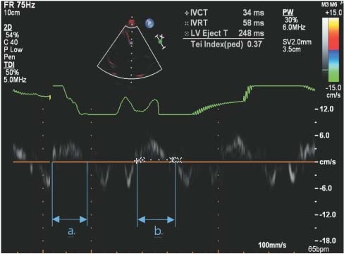
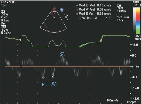
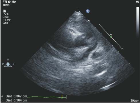
RCA, right coronary artery; LCA, left coronary artery; EF, ejection fraction; FS, fractional shortening; TDI, tissue Doppler imaging; E', early diastolic myocardial velocity; A', late diastolic myocardial velocity; S', systolic myocardial velocity; IVCT, isovolumetric contraction time; IVRT, isovolumetric relaxation time; LVET, left ventricle ejection time.

| Clinical characteristics | Group 1 (n=62) | Group 2 (n=51) | P-value |
|---|---|---|---|
| Boys | 37 (59.7) | 28 (54.9) | 0.609 |
| Girls | 25 (40.3) | 23 (45.1) | 0.703 |
| Age (year) | 4 (2–4) | 3 (1–4) | 0.045 |
| Fever duration (>5 days) | 38 (61.3) | 26 (51.0) | 0.271 |
| Conjunctival injection | 39 (62.9) | 16 (31.4) | 0.001 |
| Cervical lymphadenopathy | 20 (32.3) | 2 (3.9) | <0.001 |
| Polymorphous skin rash | 22 (35.5) | 1 (2.0) | <0.001 |
| Abnormalities of lip or oral mucosa | 19 (30.6) | 2 (3.9) | <0.001 |
| Abnormalities of extremities | 8 (12.9) | 1 (2.0) | 0.039 |
| Laboratory data | Group 1 (n=62) | Group 2 (n=51) | P-value |
|---|---|---|---|
| Hb (g/dL) | 11.4 (11.0–12.1) | 11.8 (11.2–12.3) | 0.116 |
| WBC (/μL) | 9,570 (7,417–12,677) | 10,030 (7,500–12,440) | 0.892 |
| Neutrophil (%) | 59.1 (49.9–65.7) | 57.0 (46.5–67.0) | 0.604 |
| Platelet (x109/L) | 230.0 (193.0–282.2) | 257.0 (215.0–302.0) | 0.044 |
| ESR (mm/hr) | 36.0 (26.0–49.5) | 31.0 (18.5–41.0) | 0.063 |
| CRP (mg/dL) | 5.65 (2.81–7.66) | 2.52 (1.09–5.12) | <0.001 |
| AST (IU/L) | 26.5 (17.5–32.3) | 32.0 (28.0–35.0) | <0.001 |
| ALT (IU/L) | 16.5 (13.0–29.3) | 15.0 (12.0–18.0) | 0.044 |
| Total protein (g/dL) | 6.5 (6.3–6.7) | 6.5 (6.1–6.7) | 0.858 |
| Albumin (g/dL) | 3.7 (3.6–3.9) | 3.8 (3.7–4.0) | 0.006 |
| NT-pro BNP (pg/mL) | 192.0 (82.5–316.5) | 144.5 (144.0–220.0) | 0.089 |
| Echo findings | Group 1 (n=62) | Group 2 (n=51) | P value |
|---|---|---|---|
| RCA (mm) | 2.30 (1.75–3.95) | 1.70 (1.30–2.00) | <0.001 |
| LCA (mm) | 1.80 (1.50–2.00) | 1.60 (1.50–1.90) | 0.067 |
| EF (%) | 69.45 (65.25–75.10) | 71.55 (65.08–76.73) | 0.392 |
| FS (%) | 38.15 (35.10–42.75) | 39.60 (35.03–44.20) | 0.423 |
| TDI | |||
| E’ (cm/sec) | 1.80 (1.50–2.00) | 9.73 (8.68–11.13) | 0.020 |
| A’ (cm/sec) | 69.45 (65.25–75.10) | 4.56 (3.86–6.52) | 0.805 |
| S’ (cm/sec) | 38.15 (35.10–42.75) | 5.47 (4.91–6.34) | 0.066 |
| Tei index | 1.80 (1.50–2.00) | 0.43 (0.38–0.47) | 0.302 |
| IVCT (msec) | 69.45 (65.25–75.10) | 66.00 (54.00–79.00) | 0.277 |
| IVRT (msec) | 38.15 (35.10–42.75) | 48.00 (41.00–58.00) | 0.834 |
| LVET (msec) | 1.80 (1.50–2.00) | 277.00 (252.50–298.00) | 0.696 |
Values are presented as number (%) or number (range). P value obtained from the Mann-Whitney test. P value obtained from the Fisher’s exact test. Group 1, KD with adenoviral infection; Group 2, adenoviral infection.
Values are expressed as the mean (range). Group 1, KD with adenoviral infection; Group 2, adenoviral infection. Hb, hemoglobin; WBC, white blood cell count; ESR, erythrocyte sedimentation rate; ALT, alanine aminotransferase; AST, aspartate aminotransferase; CRP, C-reactive protein; NT-proBNP, N-terminal pro-brain natriuretic peptide; RCA, right coronary artery.
Values are expressed as the mean (range). Group 1, KD with adenoviral infection; Group 2, adenoviral infection. RCA, right coronary artery; LCA, left coronary artery; EF, ejection fraction; FS, fractional shortening; TDI, tissue Doppler imaging; E', early diastolic myocardial velocity; A', late diastolic myocardial velocity; S', systolic myocardial velocity; IVCT, isovolumetric contraction time; IVRT, isovolumetric relaxation time; LVET, left ventricle ejection time.

