Abstract
Pneumocephalus is a rare complication of neuraxial procedures and is usually associated with inadvertent dural puncture or the use of air during epidural space identification. Epiduroscopic epidural neuroplasty (EEN) is performed without air injection and permits direct visualization of the epidural space; therefore, pneumocephalus after this procedure is extremely uncommon. A 71-year-old woman with a history of lumbar spine surgery underwent EEN via a caudal approach without sedation. No dural puncture was identified during the procedure. Approximately 12 hours later, she developed a non-orthostatic headache. Brain computed tomography revealed pneumocephalus in the basal cistern and left lateral ventricle. Her symptoms improved with oxygen therapy but worsened 5 days later despite radiologic resolution of the pneumocephalus. Suspected cerebrospinal fluid (CSF) leakage was treated with an epidural blood patch, which resolved her symptoms. Pneumocephalus may occur after EEN without clinically or endoscopically recognized dural injury and may coexist with CSF leakage; an occult microdural defect cannot be excluded as the underlying mechanism. Prompt imaging should be considered in patients who develop early or atypical postprocedural headache.
-
Keywords: Pneumocephalus; Neuroplasty; Epidural space; Cerebrospinal fluid leakage; Epidural blood patch; Case reports
Introduction
Pneumocephalus is a rare but recognized complication of neuraxial procedures and is most often associated with inadvertent dural puncture or the use of air during epidural space identification with the loss-of-resistance technique. Most reported cases have occurred after epidural anesthesia or epidural steroid injection when air was introduced into the epidural space [
1-
6].
Epiduroscopic epidural neuroplasty (EEN) differs from conventional epidural techniques because it is typically performed without air injection and permits direct visualization of the epidural space during catheter manipulation and adhesiolysis [
7]. Accordingly, pneumocephalus after EEN is considered extremely uncommon, particularly when no dural puncture is recognized.
Only a limited number of reports have described pneumocephalus after epiduroscopic procedures [
8], and most previously reported cases involved identifiable procedural factors, such as suspected dural violation or air entry during epidural space localization.
Here, we report a case of early-onset headache associated with intracranial pneumocephalus after EEN in a patient with multiple prior lumbar spine surgeries. No dural puncture was identified during epiduroscopic or fluoroscopic evaluation, and the patient subsequently required an epidural blood patch, suggesting concurrent cerebrospinal fluid (CSF) leakage.
Case presentation
Ethics statement
Informed consent for publication was obtained from the patient.
Patient information
A 71-year-old woman with a medical history of hypertension, diabetes mellitus, and thyroid cancer presented with chronic low back pain and persistent bilateral radicular symptoms that were refractory to conservative treatment. She had undergone 3 prior lumbar spine surgeries, the most recent of which had been performed 2 years earlier.
Clinical findings
Lumbar spine magnetic resonance imaging showed postoperative changes after posterior decompression at L4–5 and lumbar scoliosis, along with multilevel degenerative changes. Diffuse disc bulging was observed at L1–2 and L3–4. At L2–3, a moderate left central-to-subarticular disc protrusion was identified, with associated left lateral recess stenosis. At L4–5, a presumed right subarticular disc herniation had decreased in size and T2 hyperintensity, with persistent right lateral recess stenosis. Facet joint arthropathy and thickening of the ligamentum flavum were also noted throughout the lumbar spine. Based on these findings and the clinical suspicion of epidural adhesions related to prior surgeries, EEN was planned.
Timeline
The procedure was performed without sedation to permit continuous communication with the patient and to monitor for signs of dural irritation. It was conducted via a caudal approach under fluoroscopic guidance. An epiduroscope was advanced into the epidural space, and bilateral adhesiolysis was performed at L3–5. Medication and saline were administered through the working channel. Saline was delivered intermittently by hand injection at approximately 1 mL/sec, with a total volume of less than 100 mL, including both intraoperative irrigation and postprocedural catheter injection, over a total procedure time of 45 minutes. No loss-of-resistance technique using air was employed, and no definite dural puncture was identified on epiduroscopic or fluoroscopic evaluation. The procedure was completed as planned, and the patient reported no headache or discomfort during the procedure.
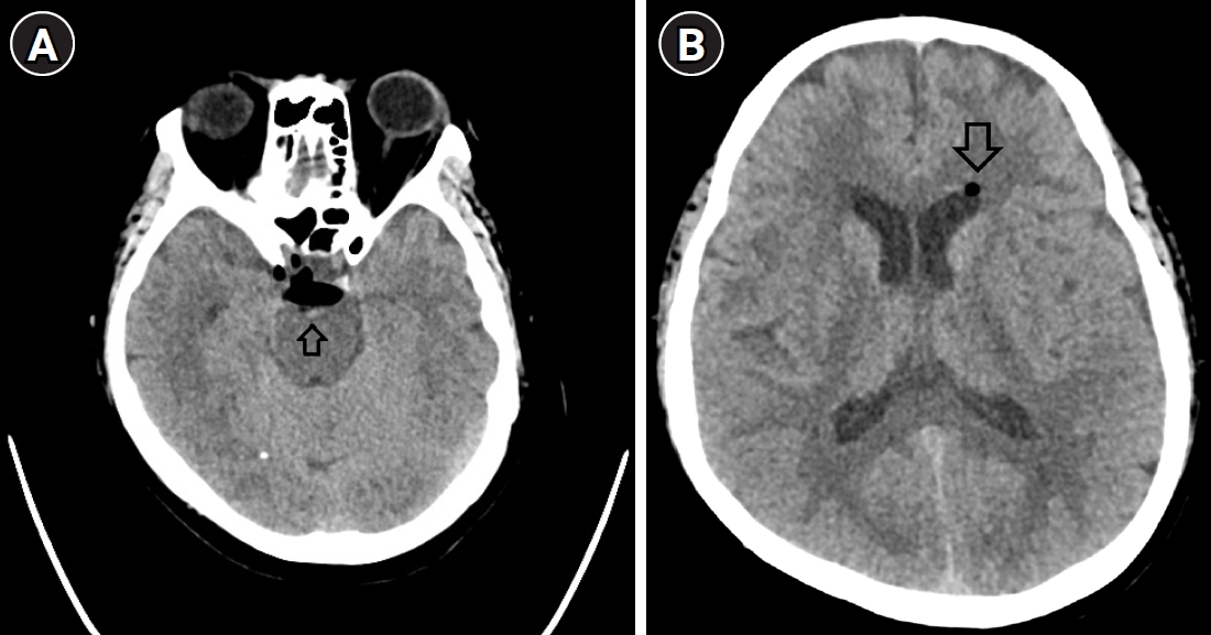
Approximately 12 hours later, the patient developed a severe, non-orthostatic headache that persisted even in the supine position. Brain computed tomography (CT) was performed to differentiate CSF leakage from other intracranial complications and revealed intracranial air in the basal cistern and left lateral ventricle, consistent with pneumocephalus (
Fig. 1). The patient was treated with supplemental oxygen, hydration, and urinary Foley catheterization for close urine-output monitoring. Her symptoms gradually improved without neurologic sequelae, and she was discharged after clinical improvement.
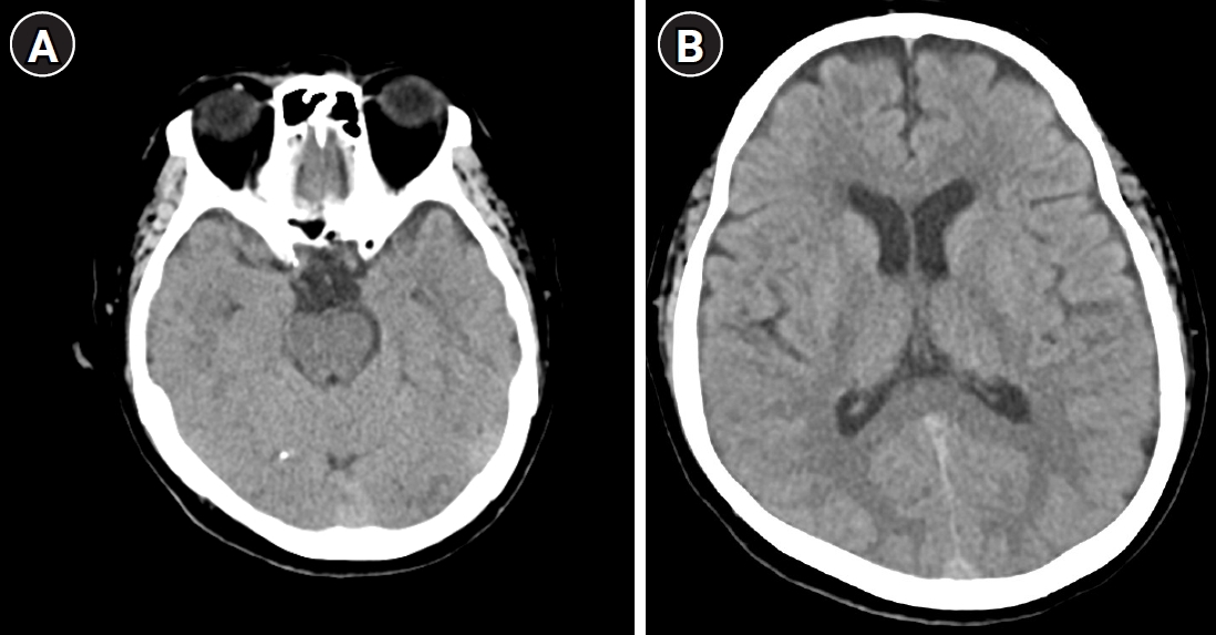
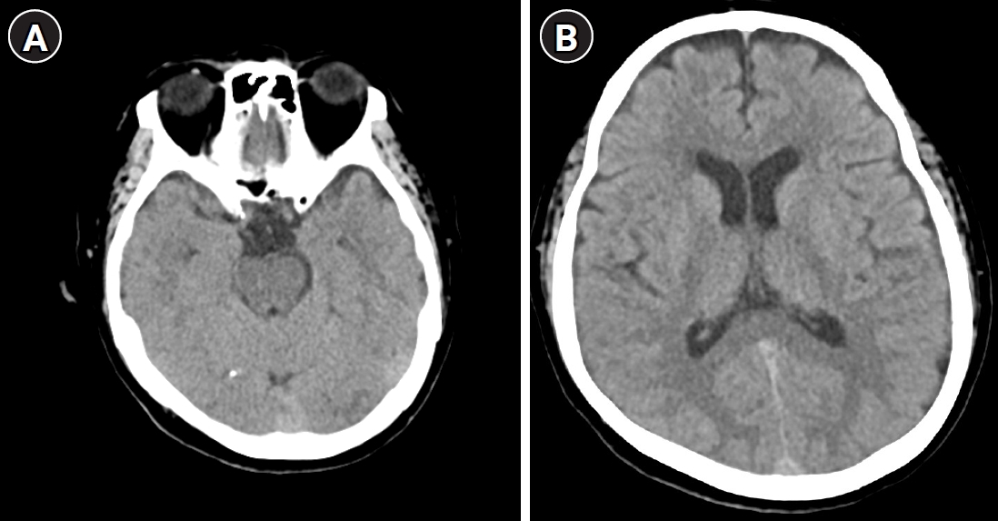
Five days after the procedure, the patient returned to the emergency department with worsening headache. The headache was diffuse, involved the entire head, and had a pressing and lancinating quality, with a numeric rating scale score of 8/10. The pain did not clearly improve in the supine position and showed no definite positional variation. Associated symptoms, including neck stiffness, nausea, tinnitus, and dizziness, were absent. Follow-up brain CT demonstrated resolution of the pneumocephalus (
Fig. 2). Conservative management was attempted initially; however, because the headache persisted and CSF leakage was suspected, an epidural blood patch was performed via a lumbar approach targeting the L5–S1 interspace with approximately 20 mL of autologous blood, resulting in symptom improvement.
Discussion
Pneumocephalus after EEN is extremely uncommon, and very few cases have been described in the published literature. To the best of our knowledge, the most relevant prior publication is a review with an accompanying case report by Marchesini et al. [
8].
A distinguishing feature of this case, compared with previous reports [
8], is the absence of sedation and intraoperative symptoms. Sedation, such as propofol, may mask patient responses to dural irritation; however, the patient remained fully awake and reported no discomfort during the procedure. This finding suggests that a microdural defect, possibly caused by increased mechanical resistance from prior surgical adhesions, may allow air migration without immediate clinical signs [
8,
9]. Although no dural puncture was identified on epiduroscopic or fluoroscopic evaluation, an occult microdural defect below the detection limits of these modalities cannot be excluded and may represent the shared pathway through which both intracranial air migration and CSF leakage occurred in this case.
The patient had undergone 3 prior lumbar spine surgeries and demonstrated multilevel degenerative changes with suspected epidural fibrosis. Extensive epidural adhesions may increase mechanical resistance during catheter manipulation and adhesiolysis, thereby increasing the risk of microscopic dural injury even when no dural puncture is visible. Previous studies have suggested that epidural fibrosis after repeated spinal surgery alters normal epidural anatomy and increases procedural difficulty during epiduroscopy, potentially predisposing patients to unintended dural microinjury [
8,
10].
Unlike previously reported cases associated with air use, the present case occurred without any air-based technique. Residual air in the epiduroscope working channel or irrigation system may have entered the subarachnoid space through an occult microdural defect that was undetectable by endoscopy or fluoroscopy. Even a small volume of air can migrate intracranially because of pressure gradients generated during saline irrigation, particularly during multilevel adhesiolysis [
8,
9]. The epiduroscope working channel and irrigation system were flushed with saline before the procedure to remove residual air; however, complete air elimination cannot be guaranteed. The use of hand-injected saline irrigation and the possibility of residual air despite preprocedural flushing are consistent with the hypothesis that even small volumes of residual air, when delivered under intermittent pressure during multilevel adhesiolysis, may be sufficient to produce intracranial pneumocephalus. Conservative management with supplemental oxygen is commonly recommended because increasing the fraction of inspired oxygen accelerates nitrogen resorption and facilitates resolution of intracranial air [
6].
Another notable feature of this case was delayed worsening of the headache several days after initial improvement. Follow-up imaging demonstrated decreased intracranial air, suggesting that pneumocephalus alone could not explain the recurrent symptoms. The recurrent headache lacked a definite orthostatic component and was not accompanied by neck stiffness, nausea, or tinnitus, which are features typically associated with intracranial hypotension. However, postural headache characteristics may be absent or atypical in intracranial hypotension [
11]. The subsequent response to an epidural blood patch strongly supports the possibility of concomitant CSF leakage with intracranial hypotension [
12]. Therefore, pneumocephalus and CSF leakage may coexist after EEN, even when no dural puncture is recognized endoscopically or fluoroscopically, and an occult microdural defect may serve as a shared pathway for both complications.
Postprocedural headache after EEN is commonly attributed to intracranial hypotension secondary to CSF leakage. However, this case demonstrates that pneumocephalus should also be considered, particularly when headache develops early or persists in the supine position [
5]. In such cases, early brain imaging may be required to differentiate pneumocephalus from post-dural puncture headache [
8].
This case also underscores the importance of close postprocedural monitoring after epiduroscopic adhesiolysis, especially in patients with multiple prior lumbar spine surgeries and suspected severe epidural fibrosis. Although EEN is generally considered safe and minimally invasive, unexpected complications, including intracranial air migration and CSF leakage, may occur even without recognized dural puncture [
8]. Therefore, short-term inpatient observation may facilitate early detection and timely management of these complications. A key limitation of this case is that CSF leakage was not objectively confirmed by imaging or direct CSF pressure measurement; the diagnosis was based on clinical suspicion and the favorable response to the epidural blood patch.
-
Authors’ contribution
Conceptualization: WK. Methodology: BKK, WK. Formal analysis: WK. Validation: WK. Project administration: SHY, WK. Writing–original draft: MJK. Writing–review & editing: WK.
-
Conflict of interest
No potential conflict of interest relevant to this article was reported.
-
Funding
None.
-
Data availability
Not applicable.
-
Acknowledgments
None.
-
Supplementary materials
None.
Fig. 1.Brain computed tomography (CT) obtained approximately 12 hours after epiduroscopic epidural neuroplasty. (A) Axial CT at the level of the basal cistern showing hypodense air collections in the interpeduncular cistern, consistent with pneumocephalus (arrow). (B) Axial CT at the level of the lateral ventricles showing a small hypodense air bubble in the anterior horn of the left lateral ventricle (arrow).
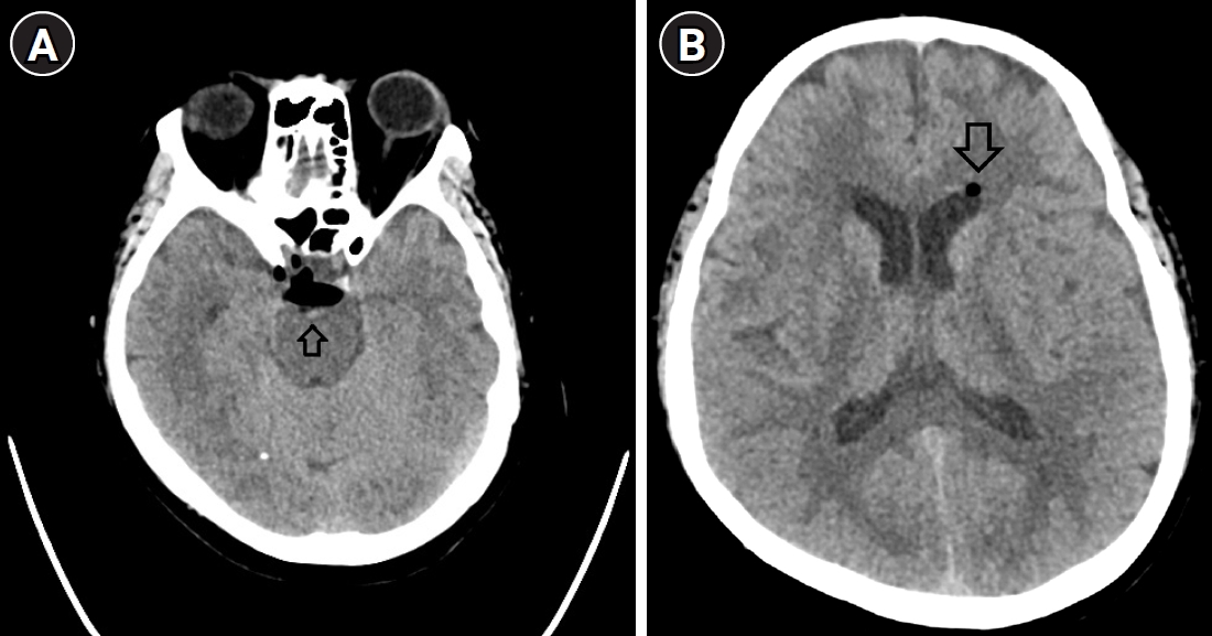
Fig. 2.Follow-up brain computed tomography (CT) obtained 5 days after the initial study. (A) Axial CT at the level of the basal cistern showing complete resolution of the previously noted pneumocephalus. (B) Axial CT at the level of the lateral ventricles showing resolution of the intraventricular air at the corresponding level.
References
- 1. Singh S, Thakur H. Pneumocephalus after lumbar epidural. Med J Armed Forces India 2018;74:397-399. https://doi.org/10.1016/j.mjafi.2017.09.011
- 2. Kim WJ, Park HG, Park YH, Shin MR, Koo GH, Shin HY. Pneumocephalus during cervical transforaminal epidural steroid injections: a case report. Am J Phys Med Rehabil 2015;94:63-69. https://doi.org/10.1097/PHM.0000000000000165
- 3. Kasai K, Osawa M. Pneumocephalus during continuous epidural block. J Anesth 2007;21:59-61. https://doi.org/10.1007/s00540-006-0446-1
- 4. Park SK, Park SH, Lee BW, Cho WJ, Choi YS. Pneumocephalus following fluoroscopy-guided lumbar epidural injection in elderly patients: two cases report and a review of Korean literatures - Two cases report -. Anesth Pain Med (Seoul) 2020;15:492-497. https://doi.org/10.17085/apm.19087
- 5. Kim YD, Lee JH, Cheong YK. Pneumocephalus in a patient with no cerebrospinal fluid leakage after lumbar epidural block: a case report. Korean J Pain 2012;25:262-266. https://doi.org/10.3344/kjp.2012.25.4.262
- 6. Castedo J, Ferreira AP, Camacho Ó. Hyperbaric oxygen therapy in the treatment of pneumocephalus associated with epidural block: case report. Braz J Anesthesiol 2021;71:295-298. https://doi.org/10.1016/j.bjane.2021.02.058
- 7. Yıldırım HU, Akbas M. Percutaneous and endoscopic adhesiolysis. Agri 2021;33:129-141. https://doi.org/10.14744/agri.2020.70037
- 8. Marchesini M, Flaviano E, Bellini V, Baciarello M, Bignami EG. Complication of epiduroscopy: a brief review and case report. Korean J Pain 2018;31:296-304. https://doi.org/10.3344/kjp.2018.31.4.296
- 9. Farshad M, Schader JF, Stauffer A, Zipser CM, Kheram N, Spirig JM, Fasser MR, Widmer J, Hagel V. Intra-, epidural and intracranial pressure changes during interlaminar endoscopy, with and without dural tear. Neurospine 2025;22:583-591. https://doi.org/10.14245/ns.2550456.228
- 10. Bosscher HA, Heavner JE. Incidence and severity of epidural fibrosis after back surgery: an endoscopic study. Pain Pract 2010;10:18-24. https://doi.org/10.1111/j.1533-2500.2009.00311.x
- 11. Kang K, Kim BK. Neurological symptoms of intracranial hypotension. J Korean Neurol Assoc 2019;37:117-122. https://doi.org/10.17340/jkna.2019.2.1
- 12. Safa-Tisseront V, Thormann F, Malassiné P, Henry M, Riou B, Coriat P, Seebacher J. Effectiveness of epidural blood patch in the management of post-dural puncture headache. Anesthesiology 2001;95:334-339. https://doi.org/10.1097/00000542-200108000-00012
Citations
Citations to this article as recorded by