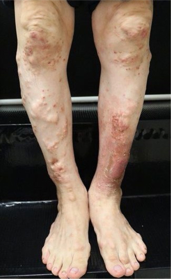
A 51-year-old man presented with multiple inflammatory skin lesions on both lower legs that had developed 3 weeks prior. He had a 15-year history of gout and stage 3 chronic kidney disease. Upon physical examination, multiple yellowish, firm papulonodules with evidence of suppuration were noted on his shins, calves, knees, and ankles (
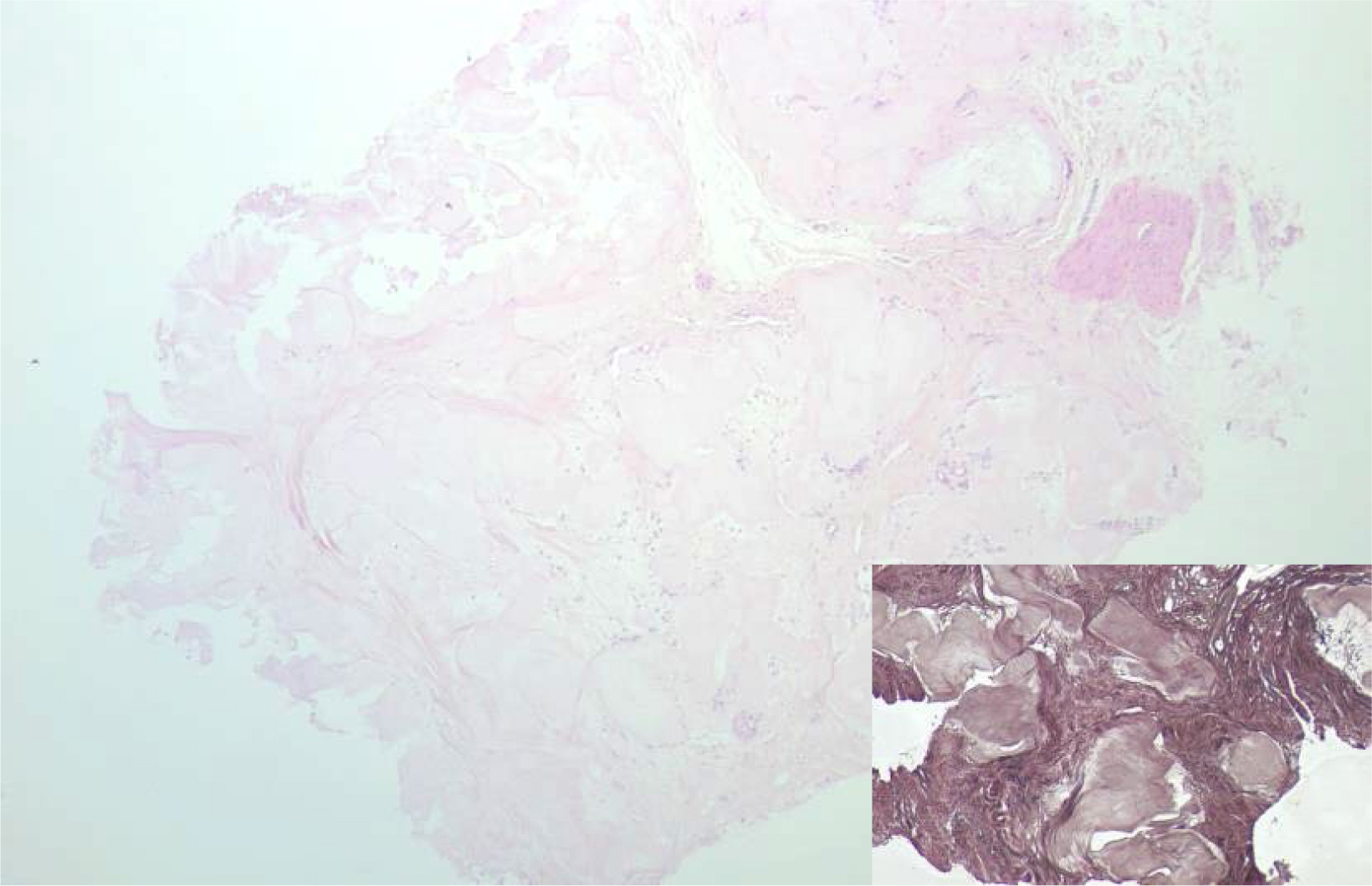
Fig. 1). A skin biopsy revealed well-circumscribed deposits of pinkish, amorphous material in the deep dermis, surrounded by histiocytic infiltration and a fibrous reaction. Von Kossa staining did not show any calcium deposition (
Fig. 2). The patient was diagnosed with disseminated cutaneous gout, and the skin lesions were treated symptomatically with topical steroids and oral antihistamines.
Fig. 1. Multiple yellowish, firm papulonodules had developed on both lower legs. Some lesions showed acute suppuration.

Fig. 2. Well-circumscribed deposition of pinkish amorphous materials was noticed in the deep dermis. Histiocytic infiltration and fibrous reaction were observed surrounding the amorphous materials (hematoxylin and eosin, ×40). Inlet, Von Kossa staining showed no calcium deposition (×100).

Gout is a chronic disease characterized by the deposition of monosodium urate (MSU) crystals, which form when urate concentrations are elevated [
1]. These MSU crystals can accumulate in joints, bones, and various body tissues, including the skin and soft tissues. The disease can be categorized into four clinical stages: asymptomatic hyperuricemia, acute gout, intercritical gout (the period between gouty attacks), and chronic tophaceous gout [
2]. Chronic tophaceous gout typically emerges after a decade or more of recurrent polyarticular gout. Gouty tophi are deposits of MSU crystals in and around joints, as well as in soft tissues. Cutaneous deposition of MSU crystals is commonly observed over joints or on the ears. Disseminated cutaneous gout is an uncommon skin manifestation of gout, characterized by widespread dermal or subcutaneous tophi that develop at extra-articular sites [
3].
The gold standard for diagnosing gout is the identification of negatively birefringent, needle-shaped MSU crystals in synovial fluid or tophi. However, the formalin fixation of skin biopsy specimens can result in the dissolution of crystals, which means that MSU deposition typically appears as pink, amorphous material in routine histological examinations. Alcohol fixation is required for the preservation and identification of the urate crystals [
4].
Ethics statement
-
Informed consent for publication of the images was obtained from the patient.
Author' Contribution
-
Conceptualization: Byun JY, Choi YW, Choi HY
Investigation: Choi YJ
Writing – original draft: Byun JY
Writing – review & editing: Choi YJ, Byun JY, Choi YW, Choi HY
Conflict of Interest
-
Ji Yeon Byun serves as the Vice Editor-in-Chief of the Ewha Medical Journal, but had no role in the decision to publish this article. No other potential conflict of interest relevant to this article was reported.
Funding
-
Not applicable.
Data availability
-
Not applicable.
Acknowledgments
Not applicable.
Supplementary materials
-
Not applicable.
References
- 1. Dalbeth N, Merriman TR, Stamp LK. Gout. Lancet 2016;388(10055):2039-2052.
- 2. Fairley JA, Aronson AB. Calcium and other mineral deposition disorders. In: In: Kang S, Amagai M, Bruckner AL, Enk AH, Margolis DJ, McMichael AJ, Orringer JS, editors. editors. Fitzpatrick’s dermatology. 9th ed. New York: McGraw Hill Education; 2019 p. p. 2313-2316.
- 3. Pradhan S, Sinha R, Sharma P, Sinha U. Atypical cutaneous presentation of chronic tophaceous gout: a case report. Indian Dermatol Online J 2010;11(2):235-238.
- 4. Guzman R, DeClerck B, Crew A, Peng D, Adler BL. Disseminated cutaneous gout: a rare manifestation of a common disease. Dermatol Online J 2020;26(1):4
Citations
Citations to this article as recorded by