1Department of Orthopaedic Surgery, School of Medicine, Kyungpook National University, Kyungpook National University Hospital, Daegu, Korea
2AIRS, Daegu, Korea
3Department of Orthopedic Surgery, St. Carolus Hospital, Faculty of Medicine, Universitas Trisakti, Jakarta, Indonesia
4Department of Orthopaedic Surgery, Gyeongsang National University Changwon Hospital, Changwon, Korea
5Department of Hand Surgery, Affiliated Hospital of Nantong University, Nantong, China
6Department of Orthopaedic Surgery, Asan Medical Center, School of Medicine, University of Ulsan, Seoul, Korea
*Corresponding author: Hyun-Joo Lee,
Department of Orthopaedic Surgery, School of Medicine, Kyungpook National
University, Kyungpook National University Hospital, 130 Donguk-ro, Jung-gu,
Daegu 41944 , Korea E-mail: lidmania@daum.net
• Received: January 1, 2024 • Revised: January 31, 2024 • Accepted: February 5, 2024
This is an Open-Access article distributed under the terms of the
Creative Commons Attribution Non-Commercial License (http://creativecommons.org/licenses/by-nc/4.0) which permits
unrestricted non-commercial use, distribution, and reproduction in any
medium, provided the original work is properly cited.
This study aimed to quantify the relationship between proximal humeral
rotation and the lateral border of the bicipital groove on fluoroscopic
imaging.
Methods:
A composite normal humerus with a marker placed on the lateral border of the
bicipital groove was affixed to a custom rotation device at the proximal cut
segment. Consecutive fluoroscopic images were captured from
−60° to 60° in 5° increments and from
−15° to 15° in 1° increments. The index value
was calculated by taking the ratio of the distance from the medial boundary
of the proximal humerus to the lateral border of the bicipital groove to the
distance between the medial and lateral boundaries of the proximal humerus.
The correlation between the humeral rotation and the index value was
determined.
Results:
The index value showed a strong positive linear correlation position during
internal rotation of the humerus across the entire range (r=0.998,
P<0.001), as well as when the humerus was externally rotated, ranging
from 15° of internal rotation to 15° of external rotation
(r=0.991, P<0.001).
Conclusion:
The lateral border of the bicipital groove may serve as a useful
intraoperative landmark for assessing proximal humeral rotation. This could
potentially enhance the outcomes of humeral fracture repair and upper arm
arthroplasty.
Restoration of the original anatomy is a crucial aspect of fracture treatment.
During surgery, coronal and sagittal angulations in long bones can be aligned
using two-dimensional (2D) fluoroscopic imaging. However, assessing rotational
deformity with conventional 2D fluoroscopic images is subjective. While the
extent of fragment rotation can be estimated by comparing the affected side to
the contralateral normal side, this method necessitates images of the
contralateral side and relies entirely on its condition. Additionally, bilateral
bone morphology is not symmetrical. In cases involving both sides, comparison is
not possible, making quantitative estimation of rotation extent unattainable.
The intraoperative three-dimensional (3D) estimation of bone position using 2D
images has garnered interest in orthopedic surgery, including computer-assisted
techniques. Rotation assessment in the lower extremity, such as the femur or
tibia, is typically performed using specific landmarks. For instance, the size
of the lesser trochanter is used as an indicator of the femur's internal
rotation status [1,2]. In contrast, studies on humeral rotation are less common
than those for the lower extremity. To date, only a handful of studies have
investigated the measurement of humeral rotation without the use of landmarks
[3–5]. Therefore, we developed a method to estimate and
evaluate the rotational alignment of the proximal humerus using a specific
landmark.
Ojbectives
The aim of the current study was to quantify the relationship between proximal
humeral rotation and the lateral border of the bicipital groove as seen on
fluoroscopic imaging. We hypothesized that the lateral border of the bicipital
groove could act as a practical landmark for assessing humeral rotation.
Methods
Ethics statement
It is not a human population study; therefore, neither approval by the
institutional review board nor obtainment of informed consent was required.
Experimental setup and acquisition of fluoroscopic images
A composite sawbone model of a humerus (#3404, Sawbones, Vashon Island, WA, USA)
was sectioned at the midpoint of the shaft. Prior to sectioning, a longitudinal
line was drawn on the anterior surface to ensure that the proximal half retained
half of this line. The proximal segment of the humeral model was then secured to
a custom rotation device, aligning the longitudinal line with the 0°
rotation mark on the device. Consequently, a 0° rotation on the device
corresponded to a neutral alignment of the proximal segment relative to the
distal segment of the humerus. For precise control and high accuracy, we
employed a modular actuator with 0.1-mm precision (Dynamixel Pro, ROBOTIS,
Seoul, Korea) as the custom rotation device. A metal dot was affixed to the
lateral edge of the bicipital groove at the point corresponding to the largest
diameter of the humeral head in preparation for fluoroscopic imaging. Since the
humeral head is spherical, any point on its surface can serve as a rotational
reference through geometric calculation. We chose the lateral edge of the
bicipital groove as this reference due to its relative ease of identification on
imaging. The location for the metal dot was specifically chosen because the
maximum circumference of the hypothetical sphere would exhibit the greatest
change with each degree of rotation, thus providing the highest sensitivity to
rotational changes. The assembly was positioned on a radiolucent table beneath
an image intensifier for imaging purposes. The rotation device was set up to
maintain the distal segment stationary while the proximal segment was rotated
from −60° to 60° in 5° increments and from
−15° to 15° in 1° increments, achieving an accuracy
of 0.1°. A fluoroscopic image was captured at each incremental position
of rotation (Fig. 1).
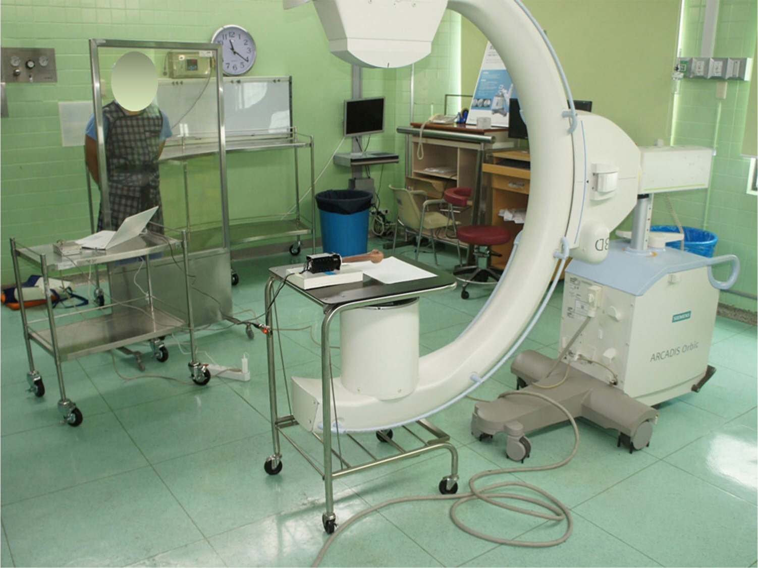
Fig. 1.
Experimental setting. The motor–humerus complex was positioned
on a radiolucent table under a C-arm. Using the rotation device, the
proximal part was rotated, while the cut distal part was fixed. A
fluoroscopic image was taken at each consecutive rotational
position.
Data analysis
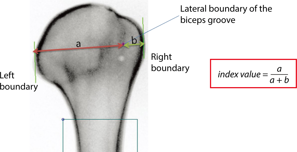
We used a specialized program to calculate an index value indicative of humeral
rotation. The user identified a rectangle and three points on fluoroscopic
images, as depicted in Fig. 2. The
rectangle was placed over the diaphysis to establish the humerus's long
axis, which was determined from the selected area through principal component
analysis. The medial and lateral boundaries of the humerus were marked, ensuring
that both were tangential to its long axis. Additionally, a point was marked at
the lateral edge of the bicipital groove. The value “a” was the
distance between the medial boundary of the humeral head, “b” was
the distance between the lateral boundary of the bicipital groove, and the value
of “a+b” was the distance from the medial to the lateral boundary.
The index value was the ratio of “a” to “a+b” in
equation 1 (Fig. 1), with the assumption
that the index value correlates with humeral rotation.
Fig. 2.
A custom program. After making a block, two lines (green lines) are
automatically generated. Lines from the medial and lateral boundary of
the humeral head to the lateral border of the bicipital groove can be
drawn perpendicularly to the green lines. The program calculated the
index value.
Statistical analysis
The Kolmogorov–Smirnov test was used to assess the normality of the
distribution. The dataset of index values followed a normal distribution;
therefore, Pearson's correlation coefficient was employed to examine the
relationship between the index value and the angle of humeral rotation. A
regression equation was also derived. The threshold for statistical significance
was established at P<0.05. Both descriptive and analytical analyses were
performed using SPSS version 15.0 (SPSS, Chicago, IL, USA).
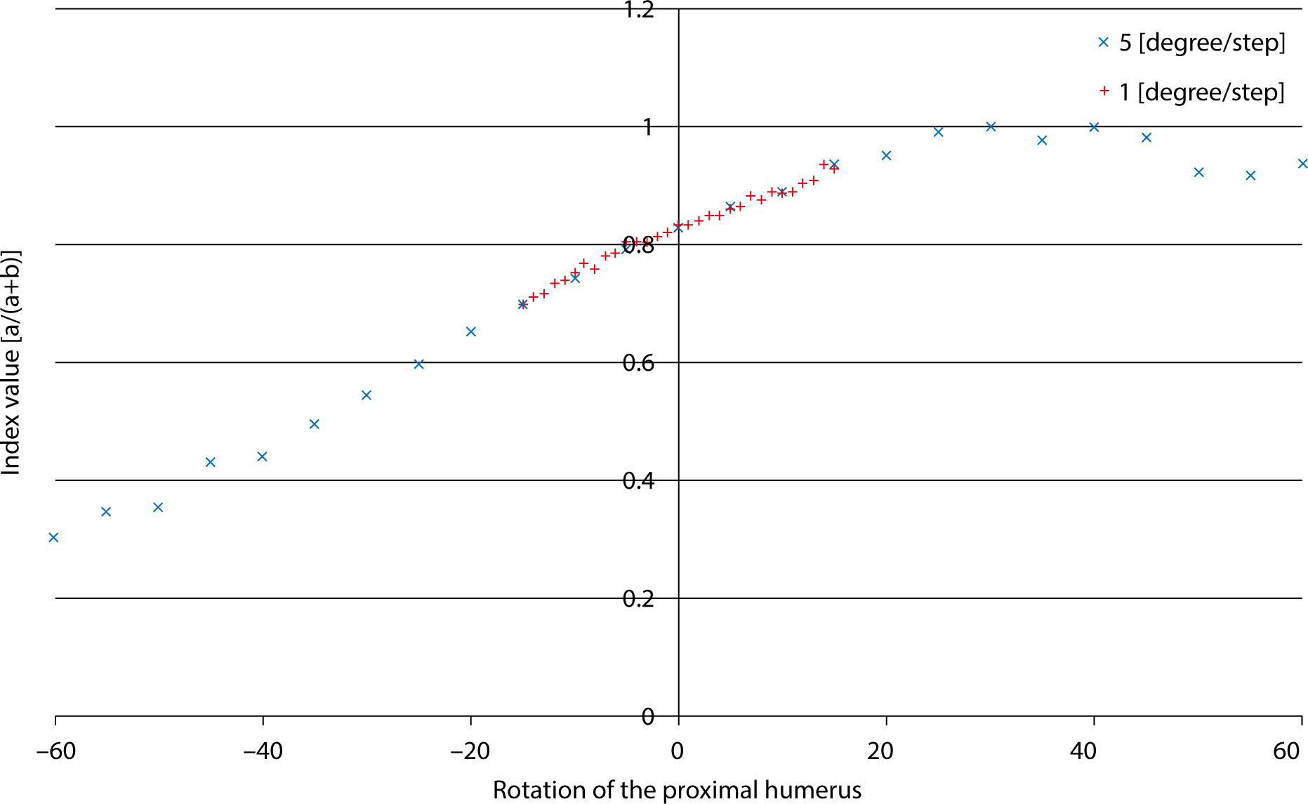
Results
The index value showed a strong positive linear correlation with position during
internal rotation of the humerus (correlation [IR]=0.998; P<0.001).
Similarly, a moderate positive linear correlation was observed with position during
external rotation of the humerus (correlation [ER]=0.693; P<0.001). Notably,
within the range of 15° internal rotation to 15° external rotation,
the correlation remained strongly positive (correlation [IR15–ER15]=0.991;
P<0.001; Fig. 3).
Fig. 3.
Linear correlation between the index value and humeral rotation. A
strongly positive correlation was observed within the range from 15°
of internal rotation to 15° of external rotation.
The regression equations for internal and external rotation were as follows:
The regression equation for internal and external rotation between −15°
and 15° was as follows:
Index value = 0.00727 × (angle) + 0.82225
Discussion
Key results
We found that the index value of the lateral border of the bicipital groove
exhibits a moderate-to-strong correlation with the rotational angle of the
humerus as seen on fluoroscopic imaging. These findings could prove beneficial
for minimally invasive plate osteosynthesis (MIPO), a technique that has
recently become more popular.
Interpretation/comparison with previous studies
MIPO offers the advantage of preserving periosteal blood supply; however, it is
often associated with rotational malalignment due to the lack of direct
visualization of the fracture site [6].
Accordingly, we established a linear correlation between the index landmark and
the rotation angle. The clinical significance of our study is that it provides a
method for estimating the rotation angle of the proximal humerus. When the
distal humerus is positioned neutrally on fluoroscopy, variations in the lateral
border of the bicipital groove can indicate the degree of rotation relative to
the distal part. This allows for the assessment of rotation without the need for
repeated fluoroscopic examination of the distal part.
The acceptable limit for rotational malalignment is generally considered to be 20
degrees. The degree of malrotation is directly related to a reduction in the
range of motion [7]. While anatomical
rotational alignment is possible using open reduction and internal fixation,
achieving correct humeral alignment during MIPO surgery can be more challenging.
In acute cases, palpating the epicondyles may be difficult due to traumatic
edema or in patients with obesity. Utilizing an index value for measurement
provides a quantitative assessment of the reduction and alignment of the
fractured fragments. Variations in humeral anatomy among different patients
necessitate this approach. By measuring the index value, surgeons can customize
the treatment to accommodate individual differences in bone structure and
alignment. This is critical for attaining optimal anatomical alignment, which is
a key factor in ensuring functional recovery.
Malrotation in the humerus is generally considered more acceptable than in the
lower extremities, which has led to limited research on humeral rotation.
Consequently, precise criteria or landmarks for assessment using plain
radiographs have yet to be established. However, studies by Itoi et al. [8] and Sabo et al. [9] have reported that humeral malrotation leads to malunion,
whereas Li et al. [4] found that it had a
negative effect on shoulder function. Moreover, recent advances in shoulder and
elbow arthroplasty have demonstrated that the sequelae of humeral malrotation
are caused by altered kinematics [8,9]. A study on humeral shaft fracture repair
assessed rotation during surgery using the cortical step sign [10]. Boileau et al. [11] used the shape of the bicipital groove to assess
rotation by comparing the ipsilateral and contralateral sides. However, neither
method was able to provide quantitative measurements. CT is highly reliable and
accurate for evaluating humeral rotation, but its feasibility during surgery is
questionable. Tan et al. [3] used the
cortical density of the lesser tuberosity as a landmark for humeral rotation and
showed its validity in a cadaver study. In contrast, our study was able to
measure rotation in 1° increments using a custom device, thus offering
superior accuracy. We utilized the lateral border of the bicipital groove as a
landmark because it provides a clear reference point when the lesser tuberosity
is not visible, particularly during ranges of internal rotation. A linear
correlation was found between the position of this landmark and the degree of
humeral rotation. This relationship may need adjustment if the proximal humerus
obscures the medial line of the greater tuberosity due to the position of the
lesser tuberosity. Nevertheless, within the clinically relevant range of humeral
rotation for computer-assisted fracture surgery (internal rotation 15° to
external rotation 15°), we observed a strong positive linear correlation
with humeral rotation. The accurate estimation of humeral rotation using a
landmark is crucial for both conventional MIPO and fracture surgery. The
bicipital groove has also been suggested as a reliable intraoperative landmark
for restoring humeral retrotorsion during shoulder replacement or for
reconstructing the premorbid anatomy of the proximal humerus [12]. Our study confirmed the reliability of
using the bicipital groove and found a linear correlation between the landmark
and the humeral rotation.
Future trends in orthopedic surgery will rely on robot-assisted or
computer-assisted techniques, which can reduce soft tissue damage and increase
the accuracy of reduction by targeting the exact point for incision and
manipulation [13]. In robot-assisted
surgery, exact data points are needed, such as for computer-assisted
arthroscopic subscapularis repair. The inability to visualize the subscapularis
tendon footprint on arthroscopy is generally accepted. However, careful
registration of the palpable lateral border of the bicipital groove allows the
surface registration of an anatomical landmark of the proximal humerus. This
improves accuracy when inserting an anchor to the lesser tuberosity. We aimed to
define accurate landmarks rather than intuitively relying on comparison with the
contralateral side. Because the anatomy of the humerus varies and a fragmented
or distorted humerus anatomy may hinder the use of this landmark, other
complementary 3D methods should be performed to determine the exact position of
the proximal humerus. The lateral border of the bicipital groove can be used as
a clinically important guide to evaluate humeral rotational alignment for
fracture reduction or other computer-assisted surgical procedures, particularly
in the range between −15° and 15°, where measurement errors
often occur.
Limitations
Our estimation method has certain limitations. First, the lateral border of the
bicipital groove may be obliterated in a comminuted fracture, severe
osteoporosis, or the presence of implant-related materials. Improperly
positioned shoulder images can also interfere with accurate imaging of the
landmark. Thus, the placement of the metal dot may not align with what is
observed in fluoroscopic images. However, the lateral border of the bicipital
groove becomes more discernible when the humerus is internally rotated. The
lesser tubercle can also serve as an intraoperative landmark for humeral
rotation. Second, we cannot generalize the data to all patients because of
variations in anatomy, such as in the degree of humeral anteversion or
anatomical variation of bicipital groove. However, our study demonstrates the
value of objective data for estimating humeral rotation, which could be used in
a practical clinical setting. Therefore, future studies are needed to determine
more generalized or normative data. Finally, estimating the rotation angle using
a shoulder image alone assumes that the elbow joint is in its neutral position,
which is relatively easy to achieve because of the wide posterior surface. Thus,
we assumed that the effect of elbow positioning is minimal.
Conclusion
The lateral border of the bicipital groove can serve as an intraoperative landmark
for the quantitative estimation of proximal humeral rotation. This landmark proves
beneficial in minimally invasive or robotic surgeries targeting the proximal
humerus. Assessing humeral rotation during surgery can enhance the results of
humeral fracture repairs and upper arm arthroplasty procedures.
Authors' contributions
Project administration: Lee HJ
Conceptualization: Lee HJ, Joung S, Tan J, Jeon IH
Methodology & data curation: Lee HJ, Joung S, Tan J
Funding acquisition: Lee HJ
Writing – original draft: Lee HJ, Joung S
Writing – review & editing: Lee HJ, Joung S, Kholinne E, Lee SJ,
Yoon JP, Tan J, Jeon IH
Conflict of interest
No potential conflict of interest relevant to this article was reported.
Funding
This work was supported by a grant from the Biomedical Research Institute,
Kyungpook National University Hospital (2015).
Data availability
Not applicable.
Acknowledgments
Not applicable.
Supplementary materials
Not applicable.
References
1. Zhang Q, Liu H, Chen W, Li X, Song Z, Pan J, et al. Radiologic measurement of lesser trochanter and its clinical
significance in Chinese. Skelet Radiol 2009;38(12):1175-1181.
2. Kim JJ, Kim E, Kim KY. Predicting the rotationally neutral state of the femur by
comparing the shape of the contralateral lesser trochanter. Orthopedics 2001;24(11):1069-1070.
3. Tan J, Lee HJ, Aminata I, Chun JM, Kekatpure AL, Jeon IH. Radiographic landmark for humeral head rotation: a new
radiographic landmark for humeral fracture fixation. Injury 2015;46(4):666-670.
4. Li Y, Wang C, Wang M, Huang L, Huang Q. Postoperative malrotation of humeral shaft fracture after plating
compared with intramedullary nailing. J Shoulder Elbow Surg 2011;20(6):947-954.
5. Park SJ, Kim E, Jeong HJ, Lee J, Park S. Prediction of the rotational state of the humerus by comparing
the contour of the contralateral bicipital groove: method for intraoperative
evaluation. Indian J Orthop 2012;46(6):675-679.
6. Wang C, Li J, Li Y, Dai G, Wang M. Is minimally invasive plating osteosynthesis for humeral shaft
fracture advantageous compared with the conventional open
technique? J Shoulder Elbow Surg 2015;24(11):1741-1748.
8. Itoi E, King GJW, Niebur GL, Morrey BF, An KN. Malrotation of the humeral component of the capitellocondylar
total elbow replacement is not the sole cause of dislocation. J Orthop Res 1994;12(5):665-671.
9. Sabo MT, Athwal GS, King GJW. Landmarks for rotational alignment of the humeral component
during elbow arthroplasty. J Bone Joint Surg 2012;94(19):1794-1800.
10. Lee HJ, Oh CW, Oh JK, Apivatthakakul T, Kim JW, Yoon JP, et al. Minimally invasive plate osteosynthesis for humeral shaft
fracture: a reproducible technique with the assistance of an external
fixator. Arch Orthop Trauma Surg 2013;133(5):649-657.
12. Vlachopoulos L, Carrillo F, Dunner C, Gerber C, Székely C, Fürnstahl P. A novel method for the approximation of humeral head retrotorsion
based on three-dimensional registration of the bicipital
groove. J Bone Joint Surg 2018;100(15):e101.
13. Micic I, Kholinne E, Hong H, Choi H, Kwak JM, Sun Y, et al. Navigation-assisted suture anchor insertion for arthroscopic
rotator cuff repair. BMC Musculoskelet Disord 2019;20(1):633
The use of the bicipital groove as an intraoperative landmark for
proximal humeral rotation during fracture fixation
Fig. 1.
Experimental setting. The motor–humerus complex was positioned
on a radiolucent table under a C-arm. Using the rotation device, the
proximal part was rotated, while the cut distal part was fixed. A
fluoroscopic image was taken at each consecutive rotational
position.
Fig. 2.
A custom program. After making a block, two lines (green lines) are
automatically generated. Lines from the medial and lateral boundary of
the humeral head to the lateral border of the bicipital groove can be
drawn perpendicularly to the green lines. The program calculated the
index value.
Fig. 3.
Linear correlation between the index value and humeral rotation. A
strongly positive correlation was observed within the range from 15°
of internal rotation to 15° of external rotation.
Fig. 1.
Fig. 2.
Fig. 3.
The use of the bicipital groove as an intraoperative landmark for
proximal humeral rotation during fracture fixation

 , Sanghyun Joung2