
| Immanuel Pradeep | 2 Articles |
|
[English]
Purpose
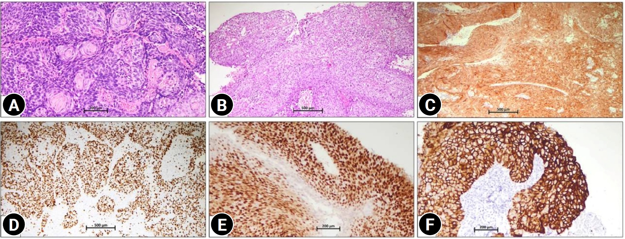
Human papillomavirus is the dominant etiological factor underlying atypical cervical squamous epithelial cell abnormalities and cervical carcinoma. Currently, only a limited number of drugs targeting specific biomarkers in cervical cancer are available. This study aimed to assess the expression of estrogen receptor (ER), human epidermal growth factor receptor 2 (HER2), and the Ki-67 proliferative index (Ki-67) in p16-positive cervical squamous premalignant and malignant lesions, which may help clarify the potential role of targeted therapies in cervical cancer. Methods: In p16-positive, histologically proven premalignant and malignant cervical lesions, ER, HER2, and Ki-67 expression were evaluated according to predefined criteria. Results: p16 showed strong nuclear and cytoplasmic positivity in 54 of 56 cases. Patchy nuclear positivity was mainly observed in low-grade squamous intraepithelial lesion (LSIL) cases (2/56). Ki-67 demonstrated a variable proliferative index ranging from 5% to 95% across all cases, with higher indices predominantly observed in squamous cell carcinomas (SCC). ER positivity in LSIL, high-grade squamous intraepithelial lesion, and SCC was 100% (2/2), 66.7% (10/15), and 46.15% (18/39), respectively. HER2 expression was predominantly negative, observed in 78.6% (44/56) of cases, equivocal in 17.8% (10/56), and positive in 3.6% (2/56). Both HER2-positive cases were SCC. ER and HER2 interpretations were analyzed and were not significantly correlated with clinical or pathological parameters. Conclusion: ER positivity decreased with progression of cervical squamous lesions, and HER2 expression was rare in cervical squamous neoplasia. No statistically significant correlation was identified between ER or HER2 expression and clinicopathological parameters. The findings of the current study may help fill gaps in the existing literature and provide essential foundational knowledge for optimizing emerging therapeutic strategies, including ER- and HER2-related therapies.
[English]
Primary leiomyosarcoma of the breast is an extremely rare malignancy, accounting for less than 1% of all breast tumors. Diagnosis is challenging because its morphology overlaps with that of other spindle cell lesions, and standardized treatment guidelines are currently unavailable. A 44-year-old woman presented with a rapidly enlarging, firm, 14.8 cm mass in the left breast. She had previously undergone surgery elsewhere, with diagnoses of leiomyoma and desmoid-type fibromatosis. Imaging demonstrated a lobulated mass without evidence of metastasis. Excision revealed pleomorphic spindle cells arranged in intersecting fascicles, with necrosis and dermal invasion. Primary breast leiomyosarcoma was confirmed by immunohistochemistry, which demonstrated smooth muscle actin and desmin positivity and negative staining for pancytokeratin, p63, S100, CD34, and BCL2. The patient underwent modified radical mastectomy followed by adjuvant radiotherapy and chemotherapy. Primary breast leiomyosarcoma is a rare entity that remains diagnostically challenging. Immunohistochemistry is essential for accurate diagnosis, and optimal management requires a dedicated multidisciplinary approach.
|
|
 , Jitendra Singh Nigam