• Contact us
  • E-Submission
ABOUT
BROWSE ARTICLES
JOURNAL POLICIES
FOR CONTRIBUTORS

Page Path

3
results for

"Min Young Lee"

Filter

Article category

Keywords

Publication year

Authors

"Min Young Lee"

Original Article

[English]
No difference in inflammatory mediator expression between mast cell-rich and mast cell-poor rosacea lesions in Korean patients: a comparative study
Jin Ju Lee, Bo Ram Kwon, Min Young Lee, Ji Yeon Byun, Joo Young Roh, Hae Young Choi, You Won Choi
Ewha Med J 2025;48(1):e78.   Published online January 31, 2025
DOI: https://doi.org/10.12771/emj.2024.e78

Objectives: This study aimed to evaluate the correlation between mast cell (MC) density in rosacea-affected skin and the expression of key inflammatory mediators, including IL-6, TNF-α, and cathelicidin LL-37. By comparing lesions rich in MCs with those having fewer MCs, we sought to elucidate the role of MCs in the inflammatory mechanisms underlying rosacea pathogenesis.

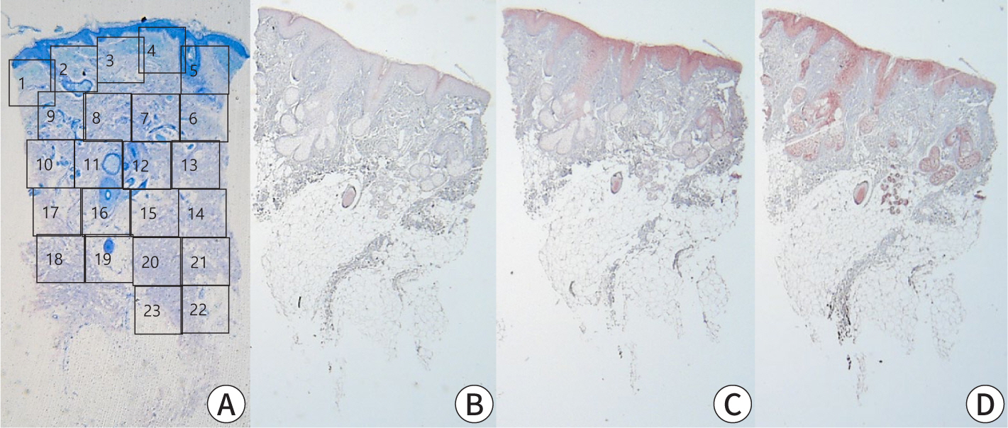
Methods: Specimens were collected from 20 patients diagnosed with rosacea who attended the outpatient clinic between 2008 and 2013. Each specimen underwent staining using hematoxylin/eosin, Giemsa, IL-6, LL-37, and TNF-α for both histopathological and immunohistochemical analyses. The number of stained cells was counted across 10 randomly selected dermal layers at a magnification of ×400 using light microscopy. The results were categorized based on the number of MCs counted: more than 10 MCs were classified as MC-rich, and 10 or fewer MCs as MC-poor.

Results: Among the 20 patients (10 MC-rich and 10 MC-poor), the MC-rich group demonstrated significantly higher MC counts than the MC-poor group (P<0.001). However, there were no significant differences in the expression levels of IL-6, LL-37, or TNF-α between the two groups. Additionally, MC density did not show any significant associations with patient demographics, clinical characteristics, or systemic comorbidities.

Conclusion: Increased MC density was not associated with differences in IL-6, TNF-α, or LL-37 expression in rosacea lesions. These findings suggest that MC infiltration may not directly influence the inflammatory mediator profile in rosacea. Further research is required to identify distinctive pathological features or markers that can elucidate the mechanisms of rosacea.

  • 88 View
  • 0 Download

Case Report

[English]
Pancreatic Panniculitis: A Case Report
Yoon Jin Choi, Min Young Lee, You Won Choi, Hae Young Choi, Ji Yeon Byun
Ewha Med J 2022;45(1):17-19.   Published online January 31, 2022
DOI: https://doi.org/10.12771/emj.2022.45.1.17

Pancreatic panniculitis is a rare skin complication in which subcutaneous fat necrosis occurs in association with pancreatic disorders, most commonly acute or chronic pancreatitis. Erythematous subcutaneous nodules develop on the legs and spontaneously ulcerate or exude an oily substance. A 32-year-old Korean female patient presented with a 2-week-history of tender nodules with erythematous crusts on her left shin. She had a history of alcoholic liver cirrhosis and, 5 weeks earlier, had been diagnosed with acute pancreatitis. The histopathologic findings from a skin biopsy were consistent with lobular panniculitis, without signs of vasculitis, and diffuse fat necrosis. Basophilic calcium deposits were present in the dermis and subcutaneous fat. These findings were suggestive of pancreatic panniculitis. The skin lesion had a chronic course corresponding to repeated exacerbations of the patient’s pancreatitis. Thus, in the differential diagnosis of subcutaneous nodules, clinicians should consider pancreatic panniculitis as a cutaneous manifestation of pancreatic disease.

  • 45 View
  • 0 Download
Images and Solution
[English]
Focal Acral Hyperkeratosis
Ji Yeon Byun, Rosa Kim, Min Young Lee, You Won Choi, Hae Young Choi
Ewha Med J 2021;44(4):144-145.   Published online October 31, 2021
DOI: https://doi.org/10.12771/emj.2021.44.4.144
  • 45 View
  • 0 Download
TOP