Clinical practice guidelines for the diagnosis and treatment of
scabies in Korea: Part 1. Epidemiology, clinical manifestations, and diagnosis
— a secondary publication
1Department of Dermatology, Jeonbuk National University Medical School, Jeonju, Korea
2Department of Dermatology, Kyung Hee University Hospital at Gangdong, Kyung Hee University School of Medicine, Seoul, Korea
3Department of Dermatology, Uijeongbu St. Mary’s Hospital, The Catholic University of Korea, Seoul, Korea
4Department of Dermatology, Incheon St. Mary’s Hospital, The Catholic University of Korea, Seoul, Korea
5Department of Dermatology, Korea University Guro Hospital, Seoul, Korea
6Department of Dermatology, Inha University Hospital, Incheon, Korea
*Corresponding author: Gwang Seong
Choi, Department of Dermatology, Inha University Hospital, 27 Inhangro, Jung-gu,
Incheon 22332, Korea, E-mail: garden@inha.ac.kr
*This is a secondary publication of Park J, Kwon SH, Lee YB, Kim HS, Jeon JH,
Choi GS. Clinical Practice Guidelines for the Diagnosis and Treatment of
Scabies in Korea: Part 1. Epidemiology, Clinical Manifestations, and
Diagnosis. Korean J Dermatol 2023;61(7):393-403 under the
permission of the editor of the Korean Journal of
Dermatology after English translation.
• Received: October 13, 2024 • Accepted: October 13, 2024
This is an Open-Access article distributed under the terms of the
Creative Commons Attribution Non-Commercial License (http://creativecommons.org/licenses/by-nc/4.0) which permits
unrestricted non-commercial use, distribution, and reproduction in any
medium, provided the original work is properly cited.
Scabies is a skin disease caused by the parasite Sarcoptes
scabiei var. hominis, which is primarily
transmitted via direct skin or sexual contact or, less commonly, via contact
with infested fomites. In Korea, the incidence of scabies has decreased from
approximately 50,000 cases per year in 2010 to about 30,000 cases per year in
2021. However, outbreaks are consistently observed in residential facilities,
such as nursing homes, especially among older adults. The clinical
manifestations of scabies vary based on the patient’s age, health status,
the number of mites, and the route of transmission. Typical symptoms of classic
scabies include intense nocturnal itching and characteristic skin rashes
(burrows and erythematous papules), with a predilection for the interdigital web
spaces, inner wrists, periumbilical areas, axillae, and genital areas. In
contrast, older adults with immunodeficiency or neurological disorders may
exhibit hyperkeratotic scaly lesions or an atypical distribution with mild to no
itching (crusted scabies). The diagnosis of scabies is based on clinical
symptoms and the results of diagnostic tests aimed at identifying the presence
of the parasite. While a history of close contact and characteristic clinical
findings suggest scabies, confirmation of the diagnosis requires detecting
scabies mites, eggs, or scybala. This can be achieved through light microscopy
of skin samples, non-invasive dermoscopy, and other high-resolution in
vivo imaging techniques.
Scabies is a highly contagious skin disease marked by severe itching, resulting
from the infestation of mites within the skin. As of 2017, over 200 million
people globally were affected, with a notably high prevalence in tropical and
low-income areas [1]. In Korea, scabies
was once prevalent, representing about 10% of outpatient visits in some general
hospitals until the early 1980s. However, the incidence declined to less than 1%
by the 1990s, likely due to strengthened public health measures. Recent years
have seen a resurgence of scabies, which is thought to be linked to the increase
in long-term care facilities associated with an aging population. In 2010, data
from the Health Insurance Review and Assessment Service reported over 50,000
cases, but this number decreased to around 30,000 cases by 2021. Despite this
decline, there remains a risk of scabies re-emerging.
To reduce scabies outbreaks, especially in communal living settings like nursing
homes, it is crucial to implement preventive measures, ensure early detection,
and adopt effective management practices. These steps help mitigate risk factors
and curb the spread of the disease. Several countries, including the U.S. and
various European nations, have established guidelines for the diagnosis,
treatment, and prevention of scabies [2–4]. In Korea, the
Korea Disease Control and Prevention Agency (KDCA) has been issuing guidance on
scabies prevention and management since 2018 [5,6]. Nevertheless, clinical
guidelines developed by dermatology experts with experience in treating scabies
are vital to broadly support healthcare providers.
Objectives
In 2023, the Korean Dermatological Association (KDA) prioritized the eradication
of scabies, launching clinical services, educational programs in communal
facilities, and public awareness campaigns. To bolster these initiatives, the
authors formed a committee within the KDA tasked with developing standardized
clinical guidelines for managing scabies. We believe this guideline will serve
as a valuable resource for everyone engaged in the treatment and prevention of
scabies.
Ethics statement
As this was a literature review study, it did not require approval from an
institutional review board. Informed consent was obtained from the patients depicted
in the figures for the use of their photographs.
Definition, transmission, and epidemiology
- Scabies is a skin infection caused by the scabies mite. It is transmitted
primarily through direct skin contact or sexual contact with an infected
individual. Transmission can also occur, though less commonly, through
indirect contact with contaminated objects.
- The number of scabies cases in Korea has steadily declined, from
approximately 50,000 in the early 2010s to around 30,000 in 2021. While
scabies is more prevalent among older adults and its incidence increases
with an aging population, about 17% of cases also occur in individuals under
the age of 20.
- Factors contributing to the ongoing occurrence of scabies cases include the
expansion of elderly care facilities, increasing drug resistance, diagnostic
difficulties due to atypical clinical presentations in clean environments or
cases of scabies incognito, and a rise in travel from regions where the
disease is endemic.
Definition
Scabies is a skin infection caused by the mite Sarcoptes
scabiei, which belongs to the phylum Arthropoda, subclass Arachnida,
order Astigmata, and family Sarcoptidae [7]. This mite parasitizes more than 40 animal species, burrowing into
the skin and causing infections in both humans and animals. There are different
varieties of scabies mites that infect humans and other animals. In Korea, three
types have been identified: S. scabiei var.
hominis (human scabies mite), S. scabiei
var. canis (dog scabies mite), and S. scabiei
var. suis (pig scabies mite). Of these, only S.
scabiei var. hominis is transmitted between humans
and causes clinical disease, and is commonly referred to as the "scabies
mite" in medical practice. Mature female mites measure 0.30–0.45
mm in length and 0.25–0.35 mm in width, while mature male mites are
approximately half that size [5]. They are
oval and grayish-white, with brown legs and a gnathosoma (mouth parts) that is
distinct from the idiosoma (body), which is short, blunt, and round with eight
legs. These mites lack eyes and respiratory organs, and feature a distinctive
long bristle on the third pair of legs.
The lifecycle of S. scabiei comprises four stages: egg, larva,
nymph, and adult [5]. After mating on the
skin surface, female mites burrow 1–2 mm into the stratum corneum. Here,
they lay an average of 35–50 eggs throughout their 4–6-week
lifespan. In contrast, male mites typically die within two days of mating. The
eggs hatch into larvae within 4–5 days, develop into nymphs, and reach
adulthood within 10–14 days. Scabies mites move at a rate of
approximately 2.5 cm per minute on the skin and can survive for 24–36
hours, and up to one week, off the host. They are most active at temperatures
above 20°C.
Transmission
Scabies primarily spreads through direct skin-to-skin or sexual contact with an
infected person. It can also spread less commonly through indirect contact with
contaminated items such as fabrics, doorknobs, bedding, or furniture.
Individuals infected with scabies can transmit the disease during the
asymptomatic incubation period and remain contagious until the mites and eggs
are eliminated through treatment. Crusted scabies, which is highly contagious,
often occurs in communal settings such as nursing homes, long-term care
facilities, prisons, and daycare centers. High-risk groups for scabies infection
include household members and sexual partners of individuals with scabies.
Crusted scabies is more likely to affect those who are immunocompromised, older
adults, individuals with physical or mental disabilities, and those in poor
conditions or with severe underlying diseases [6].
Epidemiology
In Korea, the prevalence of scabies among outpatients at eight general hospitals
across six regions, including Seoul, was approximately 2% in the 1960s, rising
to 3%–7% in the 1970s, and reaching 10% in the early 1980s. However, by
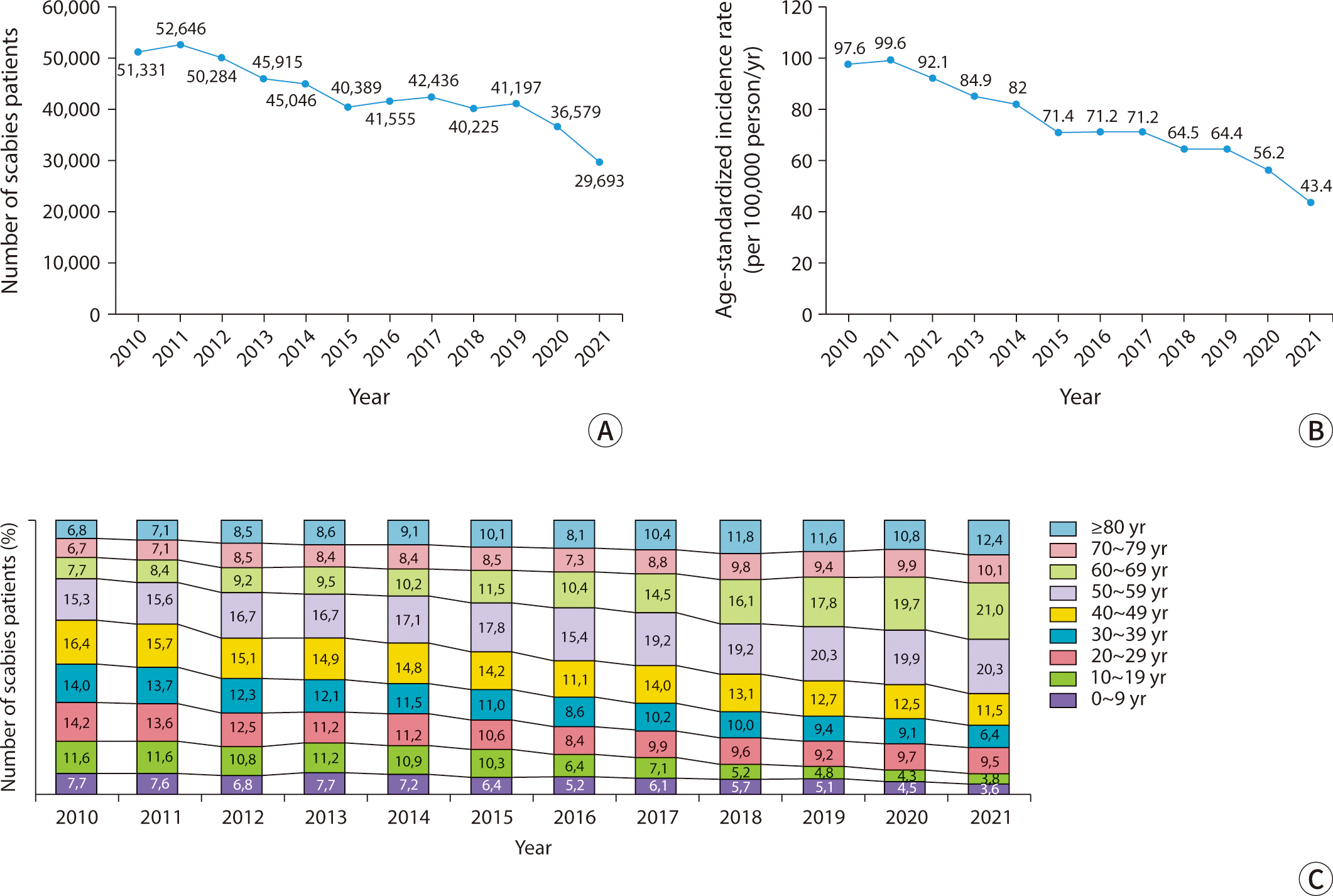
the 1990s, it had declined to below 1% [8]. Data from the Korean Health Insurance Review and Assessment Service
show that the number of scabies cases in Korea decreased from 51,331 in 2010 to
29,693 in 2021 (Fig. 1) [9]. During the same period, the
age-standardized incidence rate per 100,000 people fell from 97.6 to 43.4.
Factors contributing to this decline include improved personal hygiene,
heightened awareness of scabies in institutional settings such as nursing
facilities, and the social isolation measures and movement restrictions
associated with the COVID-19 pandemic.
Fig. 1.
Epidemiology of scabies in Korea, 2010–2021. (A) Incidence
rate. (B) Age-standardized incidence rate. (C) Age distribution of
patients.
Scabies was most prevalent among individuals in their 40s, accounting for 16% of
cases in 2010–2011. However, from 2012 to 2020, the highest incidence
shifted to those in their 50s, with a steady increase in this age group. The
higher incidence among older adults can be attributed to factors such as
population aging, an increase in long-term care facilities, and relative poverty
among older adults [9]. In contrast, among
individuals under 20, the rate of scabies declined from 33.5% of all cases in
2010 to 16.9% in 2021. The rise in scabies cases among older adults suggests
that infections in younger individuals are likely due to secondary transmission
from caregivers, healthcare workers, or family members.
The persistent incidence of scabies can be attributed to several factors,
including the expansion of elderly care facilities, increasing drug resistance,
challenges in diagnosing atypical or cryptic scabies in clean environments, and
increased travel to endemic regions [5,10]. Additionally, the
rising trend of group infections in facilities, driven by an aging population
and prolonged stays, has emerged as a significant social concern.
Clinical manifestations
- The clinical presentation of scabies varies depending on the
patient's age, health status, mite load, and route of
transmission.
- Characteristic skin symptoms of the condition include intense itching that
worsens at night, along with the presence of burrows and red bumps in
typical areas. These areas often include the interdigital web spaces of the
fingers, the inner wrists, and around the navel.
- In older adults, immunocompromised persons, or infants, scabies may
manifest differently, exhibiting less itching and atypical skin features.
These can include involvement of the scalp, face, palms, and soles, as well
as hyperkeratotic scaling or nodules.
The clinical manifestations of scabies vary based on factors such as the
patient's age, health status, underlying diseases, the number of mites
present, the route of transmission, and the time elapsed before diagnosis. While
nocturnal itching and the appearance of burrows are common symptoms, infants and
older patients with underlying conditions may present with unique and varied
symptoms. Many patients experience severe itching that disrupts sleep and diminishes
their quality of life [11]. Additionally,
they may develop skin complications such as bacterial infections, eczema, or bullous
pemphigoid [12]. In rare cases, systemic
complications such as glomerulonephritis, vasculitis, lymphadenopathy, and sepsis
may arise [13]. Scabies is typically
categorized into three types based on its clinical features: classic, crusted, and
nodular (Table 1).
Table 1.
Clinical characteristics of scabies
Classic scabies
Crusted scabies
Nodular scabies
Prevalence
Common
Rare
Uncommon (10%–30%)
Infectivity
Small number of mature mites per patient
(five or fewer in mature female mites in half of the cases)
Large numbers of mature mites per patient
(1–2 million; infectivity: very high)
Similar to classic scabies
Route of transmission
Direct skin contact
Direct skin contact
Direct skin contact
High-risk groups
Immunocompetent individuals (more common
in older adults)
Immunocompromised individuals (e.g., AIDS,
malignant tumor, autoimmune disease, immunosuppressant or steroid
use, Down syndrome, neurologic or psychological disorders)
Sexually active individuals Infants
and younger children
Pruritus
Intense (worse at night)
Mild or absent
Intense
Typical skin rash presentation
Multiple burrows, or erythematous macules
and papules (2–3 mm) (often excoriated)
Hyperkeratotic, fissured, scaly
erythematous patch or plaque (reminiscent of psoriasis or seborrheic
dermatitis)
Multiple red-brown nodules (5–20
mm) Burrows
Typical skin rash distribution
Common: fingers (interdigital web spaces),
inner wrists, extensor aspects of the extremities (elbows and
knees), periumbilical area, axillary folds, waist, buttocks, and
genital area Head, palms, and soles in infants and young
children
Bony prominences (fingers, elbows, and
iliac crest), palms and soles, head (face and scalp), auricular
region
Genital area (penis and scrotum in men),
buttock, inguinal, and axillary regions
Complications
Secondary eczema or bacterial infection,
glomerulonephritis
Erythroderma, lymphadenopathy, sepsis
AIDS, acquired immune deficiency syndrome.
Classic scabies
The primary symptom of classic scabies is intense itching that worsens at night.
An analysis of Korean scabies patients revealed that 76.4% experienced nocturnal
itching, 23.6% reported severe itching, and 13.8% had sleep disturbances due to
the intense itching. This itching is caused by the direct activity of the mites
as well as an immunological reaction, primarily Type IV delayed
hypersensitivity, to their digestive secretions, excretions, and eggs [14,15]. The worsening of symptoms at night is linked to increased mite
activity, elevated secretion levels, and heightened nerve sensitivity, which
occurs due to a decrease in sympathetic nervous activity [14].
The incubation period before the onset of itching typically ranges from 4 to 6
weeks. However, it can be shorter (i.e., less than 4 weeks) when a large number
of scabies mites are present [16]. In
cases of reinfection, symptoms can manifest within 1 to 3 days [17]. Itching may continue for several weeks
even after the mites have been eradicated. The severity of the itching often
correlates with the number of scabies lesions.
The burrows created by female mites as they tunnel through the stratum corneum
are distinctive dermatological manifestations. These burrows typically measure
2–10 mm in length and appear as grayish-white or light brown linear
tracks, either thread-like or wavy. Upon close examination, one might notice
fine scales on their surface, accompanied by slightly darker or raised edges and
ends (Fig. 2A, B) [3]. Commonly, these burrows are located between the
interdigital web spaces of the fingers, on the inner wrists, around the navel,
buttocks, male genitalia, female breasts, and armpits [2,4]. This
distribution pattern indicates the mites' preference for warmer, thinner,
and less hairy skin [18]. In
approximately 80% of patients, 1 to 4 burrows are found within the same area of
skin. Small, erythematous macules and papules, typically 2–3 mm in size
and often accompanied by severe itching, are common skin lesions resulting from
hypersensitivity to mites (Fig. 2C, D).
These lesions are usually observed around the lower abdomen, inner thighs,
axillae, and inner arms, although they do not always correspond with the
distribution of the burrows [2,17]. In most healthy adults, the scalp,
face, palms, and soles are typically unaffected. However, in infants and older
adults, one may observe burrows, papules, vesicles, and pustules on the palms
and soles [19]. In cases of prolonged
infestation, repeated scratching and infection can result in atypical
presentations, including excoriations, oozing, and crusted lesions (Fig. 2E, F).
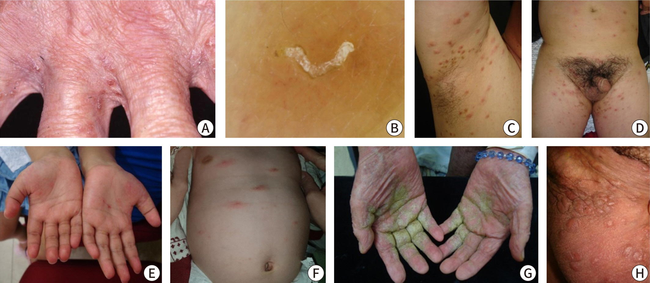
Fig. 2.
Clinical photographs of scabies. (A,B) Characteristic classic scabies
showing burrows in the interdigital web space. (C,D) Typical
erythematous macules, papules, and excoriated crusts in the typical
distribution (axilla, groin) of classic scabies. (E,F) Atypical clinical
features of scabies in infants showing palmoplantar involvement or
atypical skin lesions, such as wheals or vesicles. (G) Crusted scabies
revealing diffuse hyperkeratotic scaly lesions in immunocompromised
individuals. (H) Nodular scabies revealing multiple nodules in the
scrotum.
Crusted scabies
Previously known as Norwegian scabies, crusted scabies primarily affects
immunocompromised individuals due to physical or mental disabilities, chronic
systemic illnesses, or prolonged steroid use [20]. However, approximately 40% of cases do not have identifiable
risk factors [21]. This form of scabies
is highly contagious because of the large number of mites present, yet patients
typically experience minimal or no itching due to a diminished cellular immune
response. It is characterized by extensive, thick hyperkeratotic scales and
fissures, which resemble psoriasis or chronic eczema (Fig. 2G). Skin lesions commonly appear on the palms, soles,
head, neck, buttocks, elbows, and knees, particularly in areas subjected to
friction. Burrows may also be present in non-hyperkeratotic areas. In some
instances, the infection can extend to the nails, causing nail deformities, or
progress to erythroderma [22–24]. Secondary bacterial infections and
lymphadenopathy are common, and patients who are immunocompromised are at an
increased risk of developing sepsis [25].
Nodular scabies
Nodular scabies is a variant of classic scabies, affecting approximately
10%–30% of all cases. It is characterized by intensely itchy, red-brown
papules and nodules ranging from 5–20 mm in size. These lesions are
typically located on the male genitalia, buttocks, groin, and axillae (Fig. 2H). In infants, nodules can also be
found on the trunk, limbs, or even across the entire body. Early in the disease,
burrows may occasionally be visible on the surface of these nodules. Even after
the mites have been eradicated, the nodules often persist for some time, with
80% resolving within three months, though some may last up to a year [26].
Uncommon variants of scabies include canine scabies and scabies incognito. Canine
scabies is caused by S. scabiei var. canis and
is transmitted from dogs or other animals. Due to host specificity, it typically
resolves spontaneously in humans within a few days and does not spread between
humans [27,28]. Scabies incognito occurs when the prolonged use of
steroids or other treatments suppresses the immune and inflammatory responses,
leading to atypical clinical features [29,30]. Instead of the usual
symptoms of itching or burrows, scabies incognito presents with widespread
eczema-like lesions, which can result in a delayed diagnosis and the potential
for ongoing transmission to others.
Diagnosis
- Scabies is diagnosed clinically through a patient's history of
contact with others who have scabies and the presence of characteristic skin
symptoms. Confirmation of the diagnosis is achieved by identifying mites
using microscopy or dermoscopy.
- A clinical diagnosis of scabies is possible when burrows are visible or
when there is a history of contact with a scabies patient, accompanied by
nodules in the genital area or characteristic small papules in typical
locations.
- Microscopic examination of skin samples provides high specificity. In
contrast, dermoscopy is a quick and convenient method that achieves accuracy
comparable to microscopy when conducted by experienced clinicians. This
technique is particularly advantageous for older patients or infants who may
have difficulty cooperating during testing.
The diagnosis of scabies primarily relies on a clinical evaluation, which includes
assessing contact history and identifying characteristic skin lesions. Confirmation
is achieved through microscopy, dermoscopy, or high-magnification imaging techniques
that detect mites [2–4,31]. In
many medical institutions in Korea, there is limited access to these mite detection
tests; thus, treatment for scabies often begins based solely on the clinical
diagnosis [32]. According to a Korean
multicenter study, approximately half of the scabies cases were diagnosed clinically
without confirmatory testing. While a patient's history and typical skin
findings are invaluable for raising suspicion and aiding in the diagnosis when
confirmatory tests are unavailable, they cannot fully replace diagnostic tests.
Special consideration should also be given to immunocompromised patients, infants,
and older adults, who may not exhibit typical clinical symptoms.
Recently, a consensus was reached by 34 global scabies experts on standardized
diagnostic criteria for scabies [2,33]. These criteria categorize the diagnosis
into three levels based on diagnostic certainty: suspected, clinical, and confirmed.
This stratification provides a valuable tool for diagnosing classic scabies in
various clinical settings [34]. The
sensitivity and specificity of these standardized criteria range from 69% to 83% and
70% to 96%, respectively [33,34]. However, these criteria may be less
appropriate for atypical cases, such as crusted scabies. A simplified version of the
criteria, presented in Table 2, is also in
use, although its accuracy still requires further validation.
- Close contact with scabies patients and
pruritus - Close contact with scabies patients and skin rash
(any type)
Clinical scabies
- Scabies burrows - Close contact
with scabies patients, pruritus, and typical skin lesions (one of
the following): - Typical erythematous papules or
vesicles in a typical distribution (including the periumbilical
area, inner thigh, buttock, axilla, and inner
forearm) - Multiple nodules in the genital area or
axilla - Multiple papules, vesicles, or pustules in
the palmoplantar distribution of an infant
Confirmed scabies
- Detection of scabies mites, eggs, or
scybala in skin samples using light microscopy -
Visualization of scabies mites, eggs, or scybala using
high-resolution imaging techniques, including dermoscopy in
vivo
1)These criteria were modified from the 2020 International Alliance for the
Control of Scabies.
2)At least one item for each stage.
Clinical diagnosis
Contact history is a crucial factor in diagnosing scabies, as it is observed in
most cases. When a patient presents with pruritic skin lesions and has a history
of contact with someone diagnosed with scabies, scabies should be suspected.
This contact history can include direct interactions with the infected
individual, such as those involving cohabitants, family members, sexual
partners, roommates, healthcare providers, or caregivers. Additionally,
individuals who have had close skin contact or direct exposure to contaminated
clothing or bedding are considered to be at high risk (Table 3) [2,5]. Therefore, it is essential to thoroughly
investigate contact history, family history, sexual relationships, and any
visits or stays in long-term care facilities during the history-taking process.
However, not all patients with scabies will have an identifiable contact
history, and in some cases, the contact may not yet have been diagnosed with
scabies. Furthermore, even with a known contact history, itching and skin
lesions may not manifest until the end of the incubation period.
Table 3.
Definition of close contact and high-risk groups for transmission of
scabies
Close contact
- Skin contact1) with an
individual diagnosed with scabies - Sexual contact with
an individual diagnosed with scabies (especially, nodular
scabies) - Brief direct contact with linens (such as
towels, clothing, and bedding) used by an individual diagnosed
with scabies (especially, crusted scabies)
High-risk groups
- Family members, housemates, and sex
partners living with an individual diagnosed with
scabies - Healthcare workers, caregivers, and inpatients
who share the living environment of an individual diagnosed with
scabies (occupational exposures) - A person who has
handled linens (towels, clothing, and bedding) of an individual
diagnosed with scabies
1)At least 5–10 minutes of close and continuous contact for
acquiring classic scabies (a simple touch, handshake, and hugs are
not generally included, except in crusted scabies).
Itching is a common symptom among patients with scabies, though it is nonspecific
and alone is not sufficient for diagnosis. This is especially pertinent in older
adults, who may experience intense itching as a result of dry skin, medications,
or psychological factors. Similarly, immunocompromised individuals or young
children might exhibit mild or no itching at all; therefore, the absence of
itching should not rule out the possibility of scabies.
Clinically, scabies is often diagnosed through the observation of burrows during
a physical examination or when a patient with a history of contact presents with
distinctive skin findings, such as nodular lesions on the male genitalia or
pruritic papules in typical locations. Burrows, a hallmark of scabies, are
commonly located between the interdigital web spaces of the fingers or on the
inner wrists. Although these thread-like lesions might be visible to the naked
eye of an experienced physician, early stages of the disease, secondary eczema,
or bacterial infections can obscure the burrows. Therefore, it is advisable to
thoroughly examine typical sites. In infants, it is also important to check the
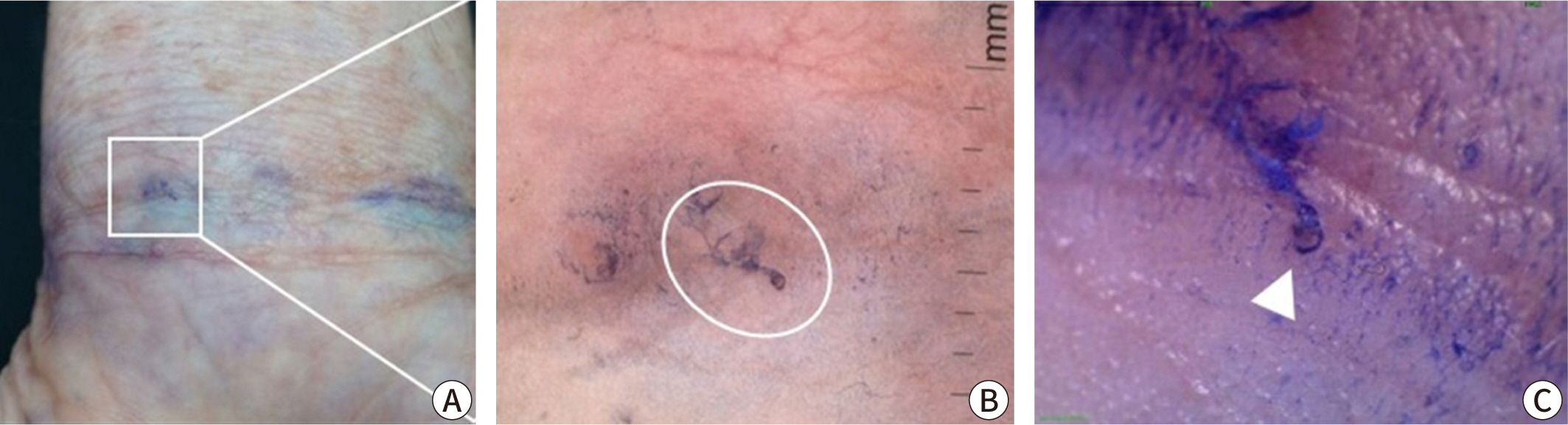
palms, soles, and scalp. The "burrow ink test" can be helpful in
identifying burrows. This test involves rubbing the suspected burrow area with a
surgical marker and then removing the excess ink with an alcohol swab (Fig. 3) [35,36]. Erythematous papules
with intense itching on the lower abdomen, inner thighs, or nodules on the male
genitalia can also support the diagnosis, although these findings are not
specific.
Fig. 3.
Burrow ink test (staining of the linear burrow with washable blue
ink). (A) Naked eye examination, (B) hand-held polarized dermoscope
(×10), and (C) high-resolution videodermoscope (×100;
circle: burrow, arrowhead: scabies mite).
Confirmed diagnosis (scabies mite detection)
Scabies can be confirmed by visually detecting live mites through a microscopic
examination of skin samples, dermoscopy, or other high-magnification imaging
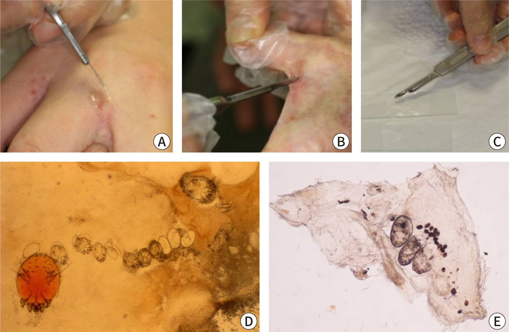
methods [2]. For microscopy, skin samples
are collected from suspected burrows using a scalpel or adhesive tape, placed on
a slide, and observed under a light microscope (Fig. 4). Applying mineral oil to the lesion before sampling helps to
capture live mites, eggs, and feces (scybala) [37]. Alternatively, potassium hydroxide can be used to dissolve the
keratin or debris in skin samples, which offers a clearer view of mites and eggs
but dissolves mite feces. Although microscopy has high specificity, it can be
cumbersome and has low sensitivity, with positive detection rates varying from
10% to 70%, depending on the examiner’s skill and sampling site [38]. Sampling from typical burrows without
abrasion of eczema on areas such as the finger webs and inner wrists can improve
detection rates. A negative result does not exclude scabies if clinical
suspicion remains high. In a Korean study, the scabies mite detection rate from
a single burrow is around 36%, and only 66.7% of scabies cases have detectable
mites [39].
Fig. 4.
Skin scraping method findings of scabies. (A–C) Skin scraping
method. Adapted from Cho [7] with
CC-BY-NC. (D,E) Microscopic findings from a skin scraping sample showing
scabies mites, eggs, and scybala in vitro (×100).
Dermoscopy is becoming increasingly popular as a quick, non-invasive, and
convenient diagnostic method. While high magnification (×50 or greater)
provides the best accuracy, skilled clinicians are able to detect mites using
handheld devices at magnifications of ×10–20 [40]. Typically, dermoscopy reveals a
distinctive brown triangular mite head, known as the "delta sign"
or "hang glider sign," along with a jet-like burrow filled with
bubbles and secretions, which gives a "jet with condensation
trail" appearance (Fig. 5) [41,42]. These features are recognized for their high sensitivity and
specificity [43–45]. Additional indicators include the wake
sign, which shows the trail of mite movement, and the gray-edge line or
bluish-white structures that represent mite feces [46]. Dermoscopy is particularly advantageous for patients
who might find sample collection distressing, such as older adults,
immunocompromised individuals, and infants, as it spares them from unnecessary
skin sample collection. However, the detection rate can vary significantly
depending on the examiner's expertise and the specific area being
observed. Other high-magnification imaging techniques, such as reflectance
confocal microscopy and optical coherence tomography, are also capable of
identifying mites within the stratum corneum, though these methods are rarely
used in clinical practice [47–50].
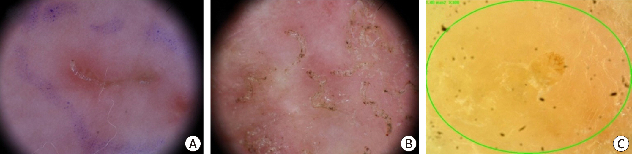
Fig. 5.
Dermoscopic findings of scabies in vivo. (A) A
triangular mite (hang glider sign) and curvilinear burrow (jet with
condensation trail; ×10). (B) Multiple scabies mites and feces
within burrows in crusted scabies (×10). (C) Scabies mite using a
high-resolution imaging device (×300).
Skin biopsies are generally not performed for diagnosing scabies; however, they
can reveal mites, eggs, and feces in the stratum corneum, as well as
inflammatory infiltrates, including eosinophils, in the dermis (Fig. 6) [51]. Blood tests do not aid in the diagnosis of scabies. While
eosinophils and immunoglobulin E levels may be elevated in patients experiencing
itchiness, these elevations are nonspecific. Tests for specific mite antibodies
are not commonly used due to their lack of sensitivity and low specificity,
which results from cross-reactivity with dust mites [52]. In cases where sexual transmission is suspected or
nodules are present in the genital area, testing for sexually transmitted
infections, such as syphilis, is advised [53].
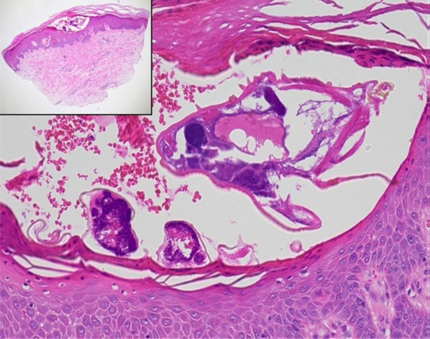
Fig. 6.
Histopathological findings of scabies showing a burrow in the stratum
corneum with Sarcoptes scabiei mites (hematoxylin and
eosin stain; ×400 [inset: ×40]).
Advanced molecular methods are being developed to detect mites. These include
nested PCR, reverse transcription-PCR targeting the cox-1 gene, matrix-assisted
laser desorption/ionization-time of flight mass spectrometry, and loop-mediated
isothermal amplification. However, further research is required to validate
these methods for routine clinical use [47,54–56].
Differential diagnosis
Given the nonspecific itching and varied clinical presentations of scabies, it is
essential to differentiate it from several other skin conditions (Table 4) [2]. The primary differential diagnoses for classic scabies include
insect bites, papular urticaria, atopic dermatitis, contact dermatitis,
folliculitis, impetigo, and pityriasis rosea. Delusional parasitosis should be
considered in patients who believe in a mite invasion on or inside the skin and
repeatedly request diagnostic testing for mite detection without a history of
contact. These patients may present objects such as hair, lint, or skin flakes,
known as "the matchbox sign," as "proof" of the
infestation, despite normal findings on examination. Crusted scabies,
characterized by its hyperkeratotic scaling, can closely resemble xerotic
eczema, psoriasis, or cutaneous T-cell lymphoma. It may also be mistaken for
seborrheic dermatitis when it appears on the head and neck. Nodular scabies may
be confused with pseudolymphoma or secondary syphilis, while common lesions on
the palms and soles in infants should be differentiated from conditions such as
infantile acropustulosis or pompholyx (dyshidrotic eczema).
Table 4.
Differential diagnosis of various types of scabies
Project administration: Park J, Kwon SH, Lee YB, Kim HS, Jeon JH, Choi GS
Conceptualization: Park J, Kwon SH, Lee YB, Kim HS, Jeon JH, Choi GS
Methodology & data curation: Park J, Kwon SH, Lee YB, Kim HS, Jeon JH,
Choi GS
Funding acquisition: not applicable
Writing – original draft: Park J, Kwon SH, Lee YB, Kim HS, Jeon JH, Choi
GS
Writing – review & editing: Park J, Kwon SH, Lee YB, Kim HS, Jeon
JH, Choi GS
Conflict of interest
No potential conflict of interest relevant to this article was reported.
Funding
Not applicable.
Data availability
Not applicable.
Acknowledgments
This study was conducted with the support of the Korean Dermatological Association.
We thank the members of the Korean Society for Cutaneous Mycology and Infection for
their valuable guidance. All images published in this paper have been used with
permission.
Supplementary materials
Not applicable.
References
1. Zhang W, Zhang Y, Luo L, Huang W, Shen X, Dong X, et al. Trends in prevalence and incidence of scabies from 1990 to 2017:
findings from the global burden of disease study 2017. Emerg Microbes Infect 2020;9(1):813-816.
2. Engelman D, Yoshizumi J, Hay RJ, Osti M, Micali G, Norton S, et al. The 2020 International Alliance for the Control of Scabies
Consensus Criteria for the diagnosis of scabies. Br J Dermatol 2020;183(5):808-820.
3. Executive Committee of Guideline for the Diagnosis and Treatment of
Scabies. Guideline for the diagnosis and treatment of scabies in Japan
(third edition). J Dermatol 2017;44(9):991-1014.
4. Salavastru CM, Chosidow O, Boffa MJ, Janier M, Tiplica GS. European guideline for the management of scabies. J Eur Acad Dermatol Venereol 2017;31(8):1248-1253.
5. Korea Disease Control and Prevention Agency. Guide for prevention and management of scabies and head lice. Cheongju: Korea Disease Control and Prevention Agency; 2018.
6. Korea Disease Control and Prevention Agency. Guide for prevention and management of scabies in convalescent
hospital. Cheongju: Korea Disease Control and Prevention Agency; 2019.
7. Cho BK. Reemerging skin disease caused by arthropods I:
scabies. J Korean Med Assoc 2011;54(5):511-520.
10. Lee SK, Kim JH, Kim MS, Lee UH. Risk factors for scabies treatment resistance: a retrospective
cohort study. J Eur Acad Dermatol Venereol 2022;36(1):126-132.
12. Romani L, Steer AC, Whitfeld MJ, Kaldor JM. Prevalence of scabies and impetigo worldwide: a systematic
review. Lancet Infect Dis 2015;15(8):960-967.
14. Kim HS, Hashimoto T, Fischer K, Bernigaud C, Chosidow O, Yosipovitch G. Scabies itch: an update on neuroimmune interactions and novel
targets. J Eur Acad Dermatol Venereol 2021;35(9):1765-1776.
20. Wong SSY, Woo PCY, Yuen K. Unusual laboratory findings in a case of Norwegian scabies
provided a clue to diagnosis. J Clin Microbiol 2005;43(5):2542-2544.
21. Roberts LJ, Huffam SE, Walton SF, Currie BJ. Crusted scabies: clinical and immunological findings in
seventy-eight patients and a review of the literature. J Infect 2005;50(5):375-381.
25. Lin S, Farber J, Lado L. A case report of crusted scabies with methicillin-resistant
Staphylococcus aureus bacteremia. J Am Geriatr Soc 2009;57(9):1713-1714.
30. Diab HM. Scabies incognito: diagnostic value of dermoscopy-guided
microscopic examination. J Egypt Women Dermatol Soc 2017;14(1):56-60.
31. Sunderkötter C, Feldmeier H, Fölster-Holst R, Geisel B, Klinke-Rehbein S, Nast A, et al. S1 guidelines on the diagnosis and treatment of scabies: short
version. J Dtsch Dermatol Ges 2016;14(11):1155-1167.
32. Park SY, Roh JY, Lee JY, Kim DW, Yoon TJ, Sim WY, et al. A clinical and epidemiological study of scabies in Korea: a
multicenter prospective study. Korean J Dermatol 2014;52(7):457-464.
33. Tsoi SK, Lake SJ, Thean LJ, Matthews A, Sokana O, Kama M, et al. Estimation of scabies prevalence using simplified criteria and
mapping procedures in three Pacific and southeast Asian
countries. BMC Public Health 2021;21(1):2060
34. Walker SL, Collinson S, Timothy J, Zayzay SK, Kollie KK, Candy N, et al. A community-based validation of the International Alliance for
the Control of Scabies Consensus Criteria by expert and non-expert examiners
in Liberia. PLoS Negl Trop Dis 2020;14(10):0008717.
38. Walter B, Heukelbach J, Fengler G, Worth C, Hengge U, Feldmeier H. Comparison of dermoscopy, skin scraping, and the adhesive tape
test for the diagnosis of scabies in a resource-poor setting. Arch Dermatol 2011;147(4):468-473.
39. Cho BK, Lee WK. Mite and tick related dermatoses. Seoul: Seoheung; 2004.
40. Lacarrubba F, Musumeci ML, Caltabiano R, Impallomeni R, West DP, Micali G. High-magnification videodermatoscopy: a new noninvasive
diagnostic tool for scabies in children. Pediatr Dermatol 2001;18(5):439-441.
41. Argenziano G, Fabbrocini G, Delfino M. Epiluminescence microscopy: a new approach to in vivo detection
of Sarcoptes scabiei. Arch Dermatol 1997;133(6):751-753.
42. Dupuy A, Dehen L, Bourrat E, Lacroix C, Benderdouche M, Dubertret L, et al. Accuracy of standard dermoscopy for diagnosing
scabies. J Am Acad Dermatol 2007;56(1):53-62.
47. Arora P, Rudnicka L, Sar-Pomian M, Wollina U, Jafferany M, Lotti T, et al. Scabies: a comprehensive review and current
perspectives. Dermatol Ther 2020;33(4):13746.
48. Lacarrubba F, Verzì AE, Micali G. Detailed analysis of in vivo reflectance confocal microscopy for
Sarcoptes scabiei hominis. Am J Med Sci 2015;350(5):414
49. Cinotti E, Labeille B, Cambazard F, Biron AC, Chol C, Leclerq A, et al. Videodermoscopy compared to reflectance confocal microscopy for
the diagnosis of scabies. J Eur Acad Dermatol Venereol 2016;30(9):1573-1577.
50. Banzhaf CA, Themstrup L, Ring HC, Welzel J, Mogensen M, Jemec GBE. In vivo imaging of Sarcoptes scabiei infestation using optical
coherence tomography. Case Rep Dermatol 2013;5(2):156-162.
51. Head ES, Macdonald EM, Ewert A, Apisarnthanarax P. Sarcoptes scabiei in histopathologic sections of skin in human
scabies. Arch Dermatol 1990;126(11):1475-1477.
54. Bae M, Kim JY, Jung J, Cha HH, Jeon NY, Lee HJ, et al. Diagnostic value of the molecular detection of Sarcoptes scabiei
from a skin scraping in patients with suspected scabies. PLoS Negl Trop Dis 2020;14(4):0008229.
55. Wong SSY, Poon RWS, Chau S, Wong SCY, To KKW, Cheng VCC, et al. Development of conventional and real-time quantitative PCR assays
for diagnosis and monitoring of scabies. J Clin Microbiol 2015;53(7):2095-2102.
56. Hahm JE, Kim CW, Kim SS. The efficacy of a nested polymerase chain reaction in detecting
the cytochrome c oxidase subunit 1 gene of Sarcoptes scabiei var. hominis
for diagnosing scabies. Br J Dermatol 2018;179(4):889-895.
Current status and epidemiology of endemic parasitic infections in Korea as of 2026: a narrative review Sun Huh Journal of the Korean Medical Association.2026; 69(3): 240. CrossRef
A case of scabies that was misdiagnosed as psoriasis Jun Ho Kwak, In Ho Bae, Hoon Choi, Chan-Ho Na, Min Sung Kim, Bong Seok Shin Journal of the Korean Society for Psoriasis.2025; 22(1): 46. CrossRef
Clinical practice guidelines for the diagnosis and treatment of
scabies in Korea: Part 1. Epidemiology, clinical manifestations, and diagnosis
— a secondary publication
Fig. 1.
Epidemiology of scabies in Korea, 2010–2021. (A) Incidence
rate. (B) Age-standardized incidence rate. (C) Age distribution of
patients.
Fig. 2.
Clinical photographs of scabies. (A,B) Characteristic classic scabies
showing burrows in the interdigital web space. (C,D) Typical
erythematous macules, papules, and excoriated crusts in the typical
distribution (axilla, groin) of classic scabies. (E,F) Atypical clinical
features of scabies in infants showing palmoplantar involvement or
atypical skin lesions, such as wheals or vesicles. (G) Crusted scabies
revealing diffuse hyperkeratotic scaly lesions in immunocompromised
individuals. (H) Nodular scabies revealing multiple nodules in the
scrotum.
Fig. 3.
Burrow ink test (staining of the linear burrow with washable blue
ink). (A) Naked eye examination, (B) hand-held polarized dermoscope
(×10), and (C) high-resolution videodermoscope (×100;
circle: burrow, arrowhead: scabies mite).
Fig. 4.
Skin scraping method findings of scabies. (A–C) Skin scraping
method. Adapted from Cho [7] with
CC-BY-NC. (D,E) Microscopic findings from a skin scraping sample showing
scabies mites, eggs, and scybala in vitro (×100).
Fig. 5.
Dermoscopic findings of scabies in vivo. (A) A
triangular mite (hang glider sign) and curvilinear burrow (jet with
condensation trail; ×10). (B) Multiple scabies mites and feces
within burrows in crusted scabies (×10). (C) Scabies mite using a
high-resolution imaging device (×300).
Fig. 6.
Histopathological findings of scabies showing a burrow in the stratum
corneum with Sarcoptes scabiei mites (hematoxylin and
eosin stain; ×400 [inset: ×40]).
Fig. 1.
Fig. 2.
Fig. 3.
Fig. 4.
Fig. 5.
Fig. 6.
Clinical practice guidelines for the diagnosis and treatment of
scabies in Korea: Part 1. Epidemiology, clinical manifestations, and diagnosis
— a secondary publication
Clinical characteristics of scabies
Classic scabies
Crusted scabies
Nodular scabies
Prevalence
Common
Rare
Uncommon (10%–30%)
Infectivity
Small number of mature mites per patient
(five or fewer in mature female mites in half of the cases)
Large numbers of mature mites per patient
(1–2 million; infectivity: very high)
Similar to classic scabies
Route of transmission
Direct skin contact
Direct skin contact
Direct skin contact
High-risk groups
Immunocompetent individuals (more common
in older adults)
Immunocompromised individuals (e.g., AIDS,
malignant tumor, autoimmune disease, immunosuppressant or steroid
use, Down syndrome, neurologic or psychological disorders)
Sexually active individuals Infants
and younger children
Pruritus
Intense (worse at night)
Mild or absent
Intense
Typical skin rash presentation
Multiple burrows, or erythematous macules
and papules (2–3 mm) (often excoriated)
Hyperkeratotic, fissured, scaly
erythematous patch or plaque (reminiscent of psoriasis or seborrheic
dermatitis)
Multiple red-brown nodules (5–20
mm) Burrows
Typical skin rash distribution
Common: fingers (interdigital web spaces),
inner wrists, extensor aspects of the extremities (elbows and
knees), periumbilical area, axillary folds, waist, buttocks, and
genital area Head, palms, and soles in infants and young
children
Bony prominences (fingers, elbows, and
iliac crest), palms and soles, head (face and scalp), auricular
region
Genital area (penis and scrotum in men),
buttock, inguinal, and axillary regions
Complications
Secondary eczema or bacterial infection,
glomerulonephritis
Erythroderma, lymphadenopathy, sepsis
AIDS, acquired immune deficiency syndrome.
Criteria for the diagnosis of scabies1)
Stage
Criteria2)
Suspected scabies
- Close contact with scabies patients and
pruritus - Close contact with scabies patients and skin rash
(any type)
Clinical scabies
- Scabies burrows - Close contact
with scabies patients, pruritus, and typical skin lesions (one of
the following): - Typical erythematous papules or
vesicles in a typical distribution (including the periumbilical
area, inner thigh, buttock, axilla, and inner
forearm) - Multiple nodules in the genital area or
axilla - Multiple papules, vesicles, or pustules in
the palmoplantar distribution of an infant
Confirmed scabies
- Detection of scabies mites, eggs, or
scybala in skin samples using light microscopy -
Visualization of scabies mites, eggs, or scybala using
high-resolution imaging techniques, including dermoscopy in
vivo
1)These criteria were modified from the 2020 International Alliance for the
Control of Scabies.
2)At least one item for each stage.
Definition of close contact and high-risk groups for transmission of
scabies
Close contact
- Skin contact1) with an
individual diagnosed with scabies - Sexual contact with
an individual diagnosed with scabies (especially, nodular
scabies) - Brief direct contact with linens (such as
towels, clothing, and bedding) used by an individual diagnosed
with scabies (especially, crusted scabies)
High-risk groups
- Family members, housemates, and sex
partners living with an individual diagnosed with
scabies - Healthcare workers, caregivers, and inpatients
who share the living environment of an individual diagnosed with
scabies (occupational exposures) - A person who has
handled linens (towels, clothing, and bedding) of an individual
diagnosed with scabies
1)At least 5–10 minutes of close and continuous contact for
acquiring classic scabies (a simple touch, handshake, and hugs are
not generally included, except in crusted scabies).
Differential diagnosis of various types of scabies
These criteria were modified from the 2020 International Alliance for the
Control of Scabies.
At least one item for each stage.
Table 3.
Definition of close contact and high-risk groups for transmission of
scabies
At least 5–10 minutes of close and continuous contact for
acquiring classic scabies (a simple touch, handshake, and hugs are
not generally included, except in crusted scabies).
Table 4.
Differential diagnosis of various types of scabies