• Contact us
  • E-Submission
ABOUT
BROWSE ARTICLES
JOURNAL POLICIES
FOR CONTRIBUTORS

Articles

Page Path

Case Report

Sessile Serrated Adenoma with High-grade Dysplasia

The Ewha Medical Journal 2012;35(1):44-48. Published online: March 31, 2012

Department of Internal Medicine, Ewha Womans University School of Medicine, Seoul, Korea.

Corresponding author: Sung-Ae Jung, Department of Internal Medicine, Ewha Womans University School of Medicine, 1071 Anyangcheon-ro, Yangcheon-gu, Seoul 158-710, Korea. Tel: 82-2-2650-5053, Fax: 82-2-2655-2076, jassa@ewha.ac.kr
• Received: August 1, 2011   • Accepted: September 2, 2011

Copyright © 2012. Ewha Womans University School of Medicine

This is an Open Access article distributed under the terms of the Creative Commons Attribution Non-Commercial License (http://creativecommons.org/licenses/by-nc/3.0/) which permits unrestricted non-commercial use, distribution, and reproduction in any medium, provided the original work is properly cited.
  • 57 Views
  • 0 Download
prev next
  • Until recently, colorectal polyps were classified predominantly as hyperplastic or adenomatous. While adenomatous polyps are well-characterized precursor lesions of adenocarcinomas, hyperplastic polyps have been considered as benign lesion. However, some hyperplastic polyps with serrated morphology of the crypts have been recognized to have distinctive features and these polyps were termed 'serrated adenomas'. Recent data show that sessile serrated adenomas (SSA) might be the precursors of serrated colonic cancers, underlining the necessity of identifying them. SSA is approximately 3% of all polyps, commonly appears as flat or sessile and yellowish due to mucus production. In the pathogenesis of SSA, progression to high grade dysplasia or early invasive carcinoma may be associated with serrated neoplasia pathway different from adenoma-carcinoma sequence. We report a case with a colon polyp diagnosed as sessile serrated adenoma with high grade dysplasia after endoscopic submucosal dissection.
  • 1. Longacre TA, Fenoglio-Preiser CM. Mixed hyperplastic adenomatous polyps/serrated adenomas: a distinct form of colorectal neoplasia. Am J Surg Pathol 1990;14:524-537.
  • 2. Fenoglio CM, Pascal RR. Colorectal adenomas and cancer: pathologic relationships. Cancer 1982;50:2601-2608.
  • 3. Makinen MJ, George SM, Jernvall P, Makela J, Vihko P, Karttunen TJ. Colorectal carcinoma associated with serrated adenoma: prevalence, histological features, and prognosis. J Pathol 2001;193:286-294.
  • 4. Aust DE, Baretton GB. Serrated polyps of the colon and rectum (hyperplastic polyps, sessile serrated adenomas, traditional serrated adenomas, and mixed polyps)-proposal for diagnostic criteria. Virchows Arch 2010;457:291-297.
  • 5. Torlakovic E, Snover DC. Serrated adenomatous polyposis in humans. Gastroenterology 1996;110:748-755.
  • 6. Goldstein NS, Bhanot P, Odish E, Hunter S. Hyperplastic-like colon polyps that preceded microsatellite-unstable adenocarcinomas. Am J Clin Pathol 2003;119:778-796.
  • 7. Leggett B, Whitehall V. Role of the serrated pathway in colorectal cancer pathogenesis. Gastroenterology 2010;138:2088-2100.
  • 8. Snover DC. Update on the serrated pathway to colorectal carcinoma. Hum Pathol 2011;42:1-10.
  • 9. Leggett BA, Devereaux B, Biden K, Searle J, Young J, Jass J. Hyperplastic polyposis: association with colorectal cancer. Am J Surg Pathol 2001;25:177-184.
  • 10. Vogelstein B, Fearon ER, Hamilton SR, Kern SE, Preisinger AC, Leppert M, et al. Genetic alterations during colorectal-tumor development. N Engl J Med 1988;319:525-532.
  • 11. Chan TL, Zhao W, Leung SY, Yuen ST. BRAF and KRAS mutations in colorectal hyperplastic polyps and serrated adenomas. Cancer Res 2003;63:4878-4881.
  • 12. Koinuma K, Shitoh K, Miyakura Y, Furukawa T, Yamashita Y, Ota J, et al. Mutations of BRAF are associated with extensive hMLH1 promoter methylation in sporadic colorectal carcinomas. Int J Cancer 2004;108:237-242.
  • 13. Iino H, Jass JR, Simms LA, Young J, Leggett B, Ajioka Y, et al. DNA microsatellite instability in hyperplastic polyps, serrated adenomas, and mixed polyps: a mild mutator pathway for colorectal cancer? J Clin Pathol 1999;52:5-9.
  • 14. Jass JR. Hyperplastic-like polyps as precursors of microsatellite-unstable colorectal cancer. Am J Clin Pathol 2003;119:773-775.
  • 15. Torlakovic E, Skovlund E, Snover DC, Torlakovic G, Nesland JM. Morphologic reappraisal of serrated colorectal polyps. Am J Surg Pathol 2003;27:65-81.
  • 16. Sheridan TB, Fenton H, Lewin MR, Burkart AL, Iacobuzio-Donahue CA, Frankel WL, et al. Sessile serrated adenomas with low- and high-grade dysplasia and early carcinomas: an immunohistochemical study of serrated lesions "caught in the act". Am J Clin Pathol 2006;126:564-571.
  • 17. Gurudu SR, Heigh RI, De Petris G, Heigh EG, Leighton JA, Pasha SF, et al. Sessile serrated adenomas: demographic, endoscopic and pathological characteristics. World J Gastroenterol 2010;16:3402-3405.
  • 18. Lu FI, van Niekerk de W, Owen D, Tha SP, Turbin DA, Webber DL. Longitudinal outcome study of sessile serrated adenomas of the colorectum: an increased risk for subsequent right-sided colorectal carcinoma. Am J Surg Pathol 2010;34:927-934.
  • 19. Snover DC. Serrated polyps of the large intestine. Semin Diagn Pathol 2005;22:301-308.
  • 20. Snover DC, Jass JR, Fenoglio-Preiser C, Batts KP. Serrated polyps of the large intestine: a morphologic and molecular review of an evolving concept. Am J Clin Pathol 2005;124:380-391.
  • 21. Bauer VP, Papaconstantinou HT. Management of serrated adenomas and hyperplastic polyps. Clin Colon Rectal Surg 2008;21:273-279.
  • 22. Noffsinger AE. Serrated polyps and colorectal cancer: new pathway to malignancy. Annu Rev Pathol 2009;4:343-364.
  • 23. Winawer SJ, Zauber AG, Fletcher RH, Stillman JS, OBrien MJ, Levin B, et al. Guidelines for colonoscopy surveillance after polypectomy: a consensus update by the US Multi-Society Task Force on Colorectal Cancer and the American Cancer Society. Gastroenterology 2006;130:1872-1885.
  • 24. Rex DK, Kahi CJ, Levin B, Smith RA, Bond JH, Brooks D, et al. Guidelines for colonoscopy surveillance after cancer resection: a consensus update by the American Cancer Society and US Multi-Society Task Force on Colorectal Cancer. CA Cancer J Clin 2006;56:160-167.
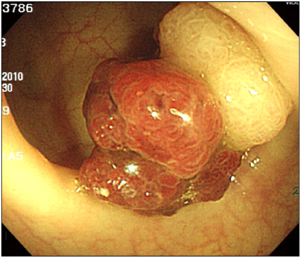
Fig. 1
The colonoscopy reveals broad-based sessile polyp of 3.3 cm in size on sigmoid colon. Its surface shows large tubular pit pattern. There is a reddish papillomatous polypoid lesion arising from the surface of the polyp.
emj-35-44-g001.jpg
Fig. 2
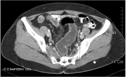
Abdominal computed tomography show no evidence of apparent wall thickening or intraluminal mass-like lesion in the colon.
emj-35-44-g002.jpg
Fig. 3
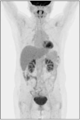
PET findings shows no abnormal hypermetabolic lesion.
emj-35-44-g003.jpg
Fig. 4
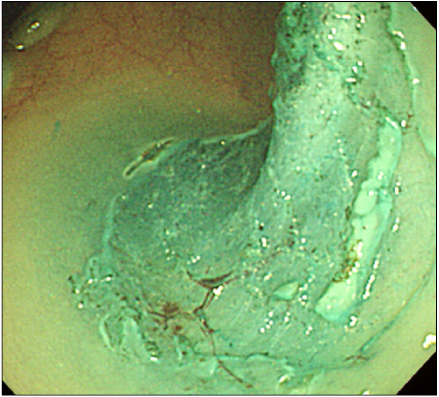
Endoscopic findings during endoscopic submucosal dissection.
emj-35-44-g004.jpg
Fig. 5
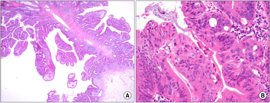
Pathologic findings. (A) The polyp is measured 3.4×2.2×2 cm in size. Its surface is granular, villous and focally had polypoid mass with long stalk (H&E stain, ×100). (B) Serrated adenoma with focal high grade dysplasia is noted with clear resection margin (H&E stain, ×200).
emj-35-44-g005.jpg

Figure & Data

References

    Citations

    Citations to this article as recorded by  

      Download Citation

      Download a citation file in RIS format that can be imported by all major citation management software, including EndNote, ProCite, RefWorks, and Reference Manager.

      Format:

      Include:

      Sessile Serrated Adenoma with High-grade Dysplasia
      Ewha Med J. 2012;35(1):44-48.   Published online March 31, 2012
      Download Citation
      Download a citation file in RIS format that can be imported by all major citation management software, including EndNote, ProCite, RefWorks, and Reference Manager.

      Format:
      • RIS — For EndNote, ProCite, RefWorks, and most other reference management software
      • BibTeX — For JabRef, BibDesk, and other BibTeX-specific software
      Include:
      • Citation for the content below
      Sessile Serrated Adenoma with High-grade Dysplasia
      Ewha Med J. 2012;35(1):44-48.   Published online March 31, 2012
      Close

      Figure

      • 0
      • 1
      • 2
      • 3
      • 4
      Sessile Serrated Adenoma with High-grade Dysplasia
      Image Image Image Image Image
      Fig. 1 The colonoscopy reveals broad-based sessile polyp of 3.3 cm in size on sigmoid colon. Its surface shows large tubular pit pattern. There is a reddish papillomatous polypoid lesion arising from the surface of the polyp.
      Fig. 2 Abdominal computed tomography show no evidence of apparent wall thickening or intraluminal mass-like lesion in the colon.
      Fig. 3 PET findings shows no abnormal hypermetabolic lesion.
      Fig. 4 Endoscopic findings during endoscopic submucosal dissection.
      Fig. 5 Pathologic findings. (A) The polyp is measured 3.4×2.2×2 cm in size. Its surface is granular, villous and focally had polypoid mass with long stalk (H&E stain, ×100). (B) Serrated adenoma with focal high grade dysplasia is noted with clear resection margin (H&E stain, ×200).
      Sessile Serrated Adenoma with High-grade Dysplasia
      TOP