Division of Cardiology, Department of Internal Medicine, Daejeon St. Mary's Hospital, The Catholic University of Korea School of Medicine, Daejeon, Korea.
Copyright © 2012. Ewha Womans University School of Medicine
This is an Open Access article distributed under the terms of the Creative Commons Attribution Non-Commercial License (http://creativecommons.org/licenses/by-nc/3.0/) which permits unrestricted non-commercial use, distribution, and reproduction in any medium, provided the original work is properly cited.


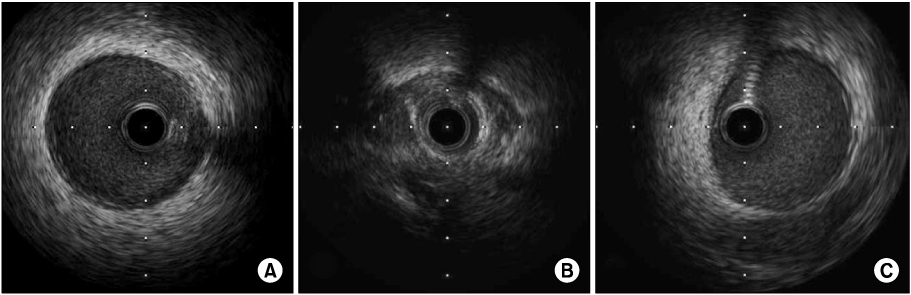
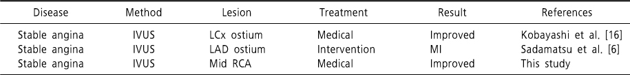
Cases of negative remodeling
IVUS, intravascular ultrasound; LCx, left circumflex artery; LAD, left anterior descending artery; RCA, right coronary artery; MI, myocardial infarction.
IVUS, intravascular ultrasound; LCx, left circumflex artery; LAD, left anterior descending artery; RCA, right coronary artery; MI, myocardial infarction.