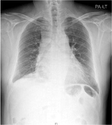
Fig. 1The initial chest X-ray shows increased interstitial markings in both lung fields, mediastinal widening and cardiomegaly.

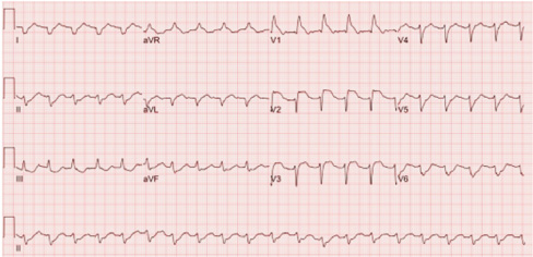
Fig. 2Electrocardiography on admission shows sinus tachycardia, right bundle branch block, ST depression in lead II, III aVF, and ST elevation in lead V1-3.

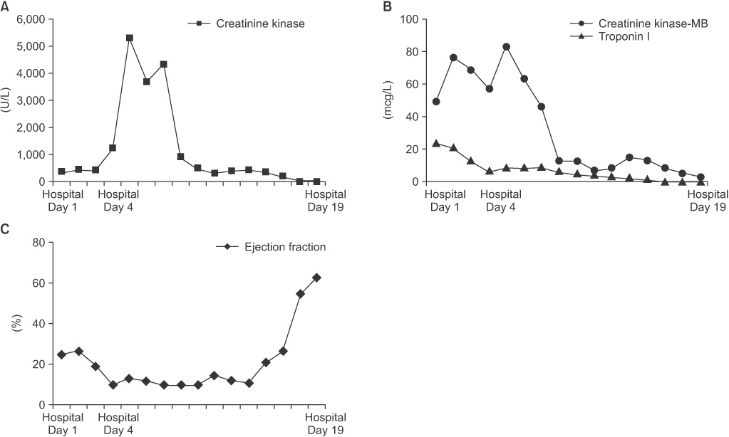
Fig. 3Changes in cardiac markers―(A) creatinine kinase, (B) creatinine kinase-MB and troponin I―and (C) ejection fraction.

Fig. 4On the 4th day of hospitalization, electrocardiography shows atrioventricular dissociation (A). After improvement, electrocardiography shows normal atrio-ventricular conduction (B).
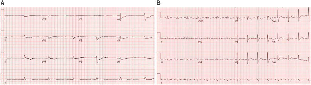
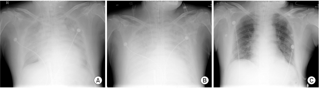
Fig. 5During the follow up period, chest X-ray shows pleural effusion and pulmonary edema and cardiomegaly, and aggravation for about a week. After three weeks of admission, clinical improvement has been observed and chest X-ray shows improvement of pleural effusion and pulmonary edema and cardiomegaly. On the (A) 4th, (B) 8th, (C) 19th day of hospitalization.