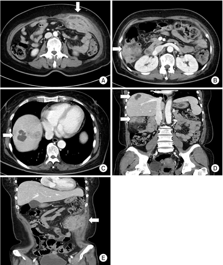
Fig. 1Abdominal computed tomography (CT). It shows 7.5 cm heterogenous enhancing soft tissue mass with necrotizing component at left lower quadrant of abdominal wall (A) (arrow), 5 cm inflammatory lesion with right paracolic gutter invasion (B) (arrow), and 3.5 cm low density lesion with peripheral rim enhancement at the right lobe of liver (C) (arrow). (D, E) Three lesions had no internal connection on coronal view (arrows).

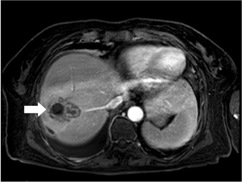
Fig. 2Liver magnetic resonance imaging. It reveals abscess with peripheral rim enhancement in right hepatic dome portion.

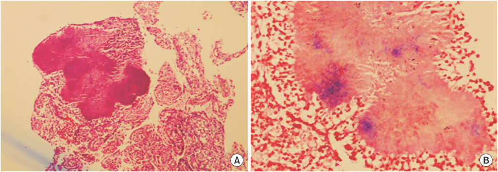
Fig. 3Histopathologic findings. (A) Sulfur granules are in the granulation tissues of abdominal wall specimen (H&E, ×100). (B) On staining with the gram stain, several colonies consist of gram-positive, branching filamentous microorganisms within the granule (Gram stain, ×400).