1Department of Surgery, Pohang Naval Hospital, Pohang, Korea
2Department of Colon and Rectal Surgery, Asan Medical Center, University of Ulsan College of Medicine, Seoul, Korea
*Corresponding author: Jong Lyul Lee,
Department of Colon and Rectal Surgery, Asan Medical Center, University of Ulsan
College of Medicine, 88 Olympic-ro 43-gil, Songpa-gu, Seoul 05505, Korea, Tel:
82-2-3010-1732, Fax: 82-2-3010-6710, E-mail:
iamleejong@amc.seoul.kr
• Received: September 30, 2022 • Accepted: October 4, 2022
This is an Open-Access article distributed under the terms of the
Creative Commons Attribution Non-Commercial License (http://creativecommons.org/licenses/by-nc/4.0) which permits
unrestricted non-commercial use, distribution, and reproduction in any
medium, provided the original work is properly cited.
Fecal incontinence (FI) is recurrent uncontrolled passage of fecal material in
patients. The life expectancy of humans has increased. Elderly patients have a
significant rate of FI. Therefore, the number of patients with FI will increase.
For diagnosis of FI, the digital rectal exam, ultrasonography, and anal
manometry are used. In addition, the severity of FI can be assessed using the FI
score system by examining symptoms. Recent applications include
three-dimensional ultrasonography and other novel approaches. The treatments for
FI include biofeedback therapy, anal implant, artificial sphincter, nerve
modulation, SECCA, stem cell therapy, and surgical intervention. Biofeedback
therapy is a noninvasive procedure. Anal implant, stem cell therapy, and SECCA
are all minimally invasive treatments. And more methods constitute intrusive
treatment. None of these therapies has been conclusively demonstrated to be
superior. Depending on the severity of the symptoms, a non-invasive approach or
an intrusive treatment is most frequently employed. In this review, I will
discuss the diagnosis and treatment options for FI.
Socially and psychologically, fecal incontinence (FI) is a tragedy. Normal bowel
continence is a complicated process including the anal sphincters, pelvic floor,
stool volume, rectal compliance, and neurologic function. It's possible for
FI to occur if even one of these things goes wrong [1].
Due to the humiliating nature of FI symptoms, many are reluctant to disclose their
illnesses to doctors. Because individuals underreported their symptoms, the true
prevalence of FI was unclear. Consequently, the prevalence of FI has fluctuated
widely between 2% and 20% [2,3]. FI prevalence varies with age. The
prevalence of FI rose with age, from 2.91% in individuals aged 20% to 29% to 16.16%
in those aged 70 and older [2]. Extremely high
incidence of FI are found among nursing home residents. The prevalence of FI is
considerably higher than 50% among nursing home residents [3,4]. Currently,
breakthroughs in sphincter-preserving surgery for rectal cancer result in
incontinence, which is a symptom of the low anterior resection syndrome [5,6].
Consequently, the old population is anticipated to grow. Consequently, the
prevalence of FI is anticipated to increase. The treatment of FI ranges from
non-invasive strategies, such as lifestyle modifications, medical pills, sphincter
augmentation with injections, and biofeedback therapy (BFT), to invasive sacral
nerve stimulation (SNS) and surgical interventions [7–9].
Given that the number of patients with FI is likely to rise, it is crucial to
identify the treatments now in development and to establish treatment indications.
In this review, we therefore considered BFT and surgical procedures for FI.
Definition and Etiology of Fecal Incontinence
Rome IV diagnostic criteria described FI as "recurrent uncontrolled flow of
feces in an individual with at least 4 years of developmental age." [10]. With the release of Rome IV diagnostic
criteria in 2016, the definition of FI was modified. Rome III separated structural
and neurogenic factors for functional FI. However, Rome IV defines FI as the
involuntary passage of solid or liquid stool, regardless of the underlying reason.
This indicates that there are several, overlapping causes of FI, and that patients
with purely psychosocial or bowel habit disorders are uncommon. Additionally, there
was no effective treatment advice based on the differentiation between causes of FI.
The second modification to the diagnostic criteria for FI concerns the definition of
event frequency. FI is defined as the development of symptoms at least six months
prior with two to four episodes of FI over four weeks. In contrast, Rome III
required only one FI event within the previous three months [11]. According to several researchers, this restricted
modification negatively impacts the quality of life for some patients. This
stringent frequency threshold resulted in a lower prevalence of FI compared to prior
estimations. These excluded patients have a diminished life quality [12]. Therefore, additional research is
necessary to provide a precise definition of FI in Rome V. Damages to the continence
process, including the anal sphincter, bowel habits, rectal compliances, pelvic
floor muscles, and rectal sensibility, might cause FI. Multiple variables cause FI
in 80% of individuals [13]. Several studies
have identified FI risk factors (Table 1)
[13–15]. Age and gastrointestinal (GI) diseases that cause stool
alterations are risk factors for FI. Aging impacts the etiology of FI, resulting in
a decrease in pelvic floor muscle mass and an increase in co-morbidities that
compromise the mechanism of continence. In addition, sphincters may be altered by
fibrosis and thickening as a result of decreased resting tone, with the external
anal sphincter's thinning resulting in a decrease in anal pressure. Moreover,
diminished rectal sensitivity, rectal compliance, and rectal capacity impede anal
sensory, motor, and rectal reservoir function in the aged [16]. Inflammatory bowel disease and irritable bowel syndrome
characterized mostly by diarrhea are risk factors for FI. However, any GI condition
that causes frequent bowel movements or a change in bowel habits can lead to FI
[17]. And the structural causes of FI are
obstetric injuries [18], anorectal surgery
[19,20], rectal prolapse [21], and
anal intercourse [22]. Puborectal or anal
sphincter dysfunction may result in FI. These injuries are possible during
spontaneous vaginal birth or forceps-assisted surgical delivery. Ischemia,
laceration, and compression of anal muscles caused by spontaneous labor and delivery
contribute to incontinence [23]. Even in the
absence of complications, up to 35% of first-time vaginal deliveries may result in
hidden sphincter injury. Risk factors for FI included forceps-assisted surgical
delivery, occipitoposterior presentation of the infant, and protracted labor [16]. Patients who underwent anorectal
procedures such as hemorrhoidectomy, sphincterotomy, and fistula surgery were
frequently discovered to have FI [24]. Rectal
prolapse results in incontinence as a result of rectal mucosa transporting feces via
the anus. And chronic rectal prolapse causes anal sphincter dilatation and
dysfunction [21].
Table 1.
Risk factors of fecal incontinence
Category
Factors
Patient
Aging, obesity, smoking, post-menopause,
nursing home residency
As with any disorder, FI requires a more thorough medical history. Patients are
hesitant to discuss their incontinence problems regularly due to their feelings of
embarrassment. The history of a patient's symptoms must be taken with care,
thoroughness, and sensitivity [25].
Clinicians should question thoroughly about the patient's underlying
condition, medications, and surgical history. In addition to obstetric and anorectal
surgery, which are directly associated, it is vital to examine the patient's
surgical history, including procedures that appear unrelated, such as
cholecystectomy and spinal surgery. Additionally, it is vital to consider drugs for
diarrhea and constipation. Changes in bowel, feces, and gas patterns must also be
evaluated. Bowel diaries and the Bristol stool form scale can help describe the
severity of FI symptoms [26].
Scoring FI systems have been utilized for objective evaluation and evaluation of
therapy outcomes. The Wexner Cleveland Clinic Score [27] and the St. Mark's Incontinence Score [28] in conjunction with the FI Quality of Life (FIQL) scale
[29] accepted by the American Society of
Colon and Rectal Surgeons (Table 2). CCF is
the most widely used historical scale. The CCF comprises of five questions, and the
sum of each score allows for the diagnosis of FI. The FI Quality of Life scale
analyzes performance across four broad categories. Lifestyle, coping/behavior,
depression/self-perception, and shame are these categories [29]. St. Mark's score is determined by four criteria:
solid stool incontinence, liquid stool incontinence, gas, and lifestyle changes
[28]. There are various scales that can
be used to assess the severity of FI symptoms. All of these measures have been
employed to diagnose FI, despite the fact that only one has been identified. The
physical examination involves a perineal skin inspection for scars from trauma,
childbirth, past surgery, fistula, or hemorrhoids, as well as a digital rectal exam.
The perineal body of female patients was palpated to see if it has been thinning.
The Valsalva technique is a strong attempt to exhale against a closed airway,
typically performed by closing the mouth and pinching the nose while exhaling. This
action is beneficial for distinguishing rectal prolapse. Examining the perianal
sensation to pinprick and the ano-cutaneous "wink" reflex will provide
a straightforward assessment of neurologic function. A digital rectal examination
can detect a fistula or hemorrhoid tumor or lesion. With the doctor's finger
inserted into the patient's anus, the patient is instructed to squeeze the
sphincter, which provides a general assessment of both resting tone and voluntary
squeeze. Colonic exams such as colonoscopy and anoscopy are also helpful to diagnose
FI [30].
Lack of ability to defer
defecation for 15 minutes
0
4
Score of 0=perfect continence;
24=totally incontinence never, 0; rarely, 1/ month;
sometimes, >1/ month but <1 a week; weekly, ≥1/
week but <1 a day; daily, ≥1/ day
Fecal incontinence quality of
life (FIQL) scale composition
Questions: scale
1-lifestyle
I cannot do many of
things I want to do. I am afraid to go
out. It is important to plan my schedule
around my bowel pattern. I cut down on how
much I eat before I go out. It is difficult
for me to get out and do things like going to a movie or to
church. I avoid traveling by plane or
train. I avoid
traveling. I avoid visiting
friends. I avoid going out to
eat. I avoid staying overnight away from
home.
Scale 2. coping/behavior
I have sex less often
than I would like to. The possibility of bowel
accidents is always on my mind. I feel I have
no control over my bowels. Whenever I go
someplace new, I specifically locate where the bathrooms
are. I worry about not being able to get to
the toilet in time. I worry about bowel
accidents. I try to prevent bowel accidents by
staying very near a bathroom. I can’t
hold my bowel movement long enough to get to the
bathroom. Whenever I am away from home, I try
to stay near a restroom as much as possible.
Question: scale
3-depression/self perception
In general, would you
say your health is excellent, very good, good, fair, and
poor. I am afraid to have
sex. I feel different from other
people. I enjoy life
less. I feel like I am not a healthy
person. I feel
depressed. During past month, have you felt so
sad, discouraged, hopeless, or had so many problems that you
wondered if anything was worthwhile.
Question: scale
4-embarrassment
I leak stool without
even knowing it. I worry about others smelling
stool on me. I feel ashamed.
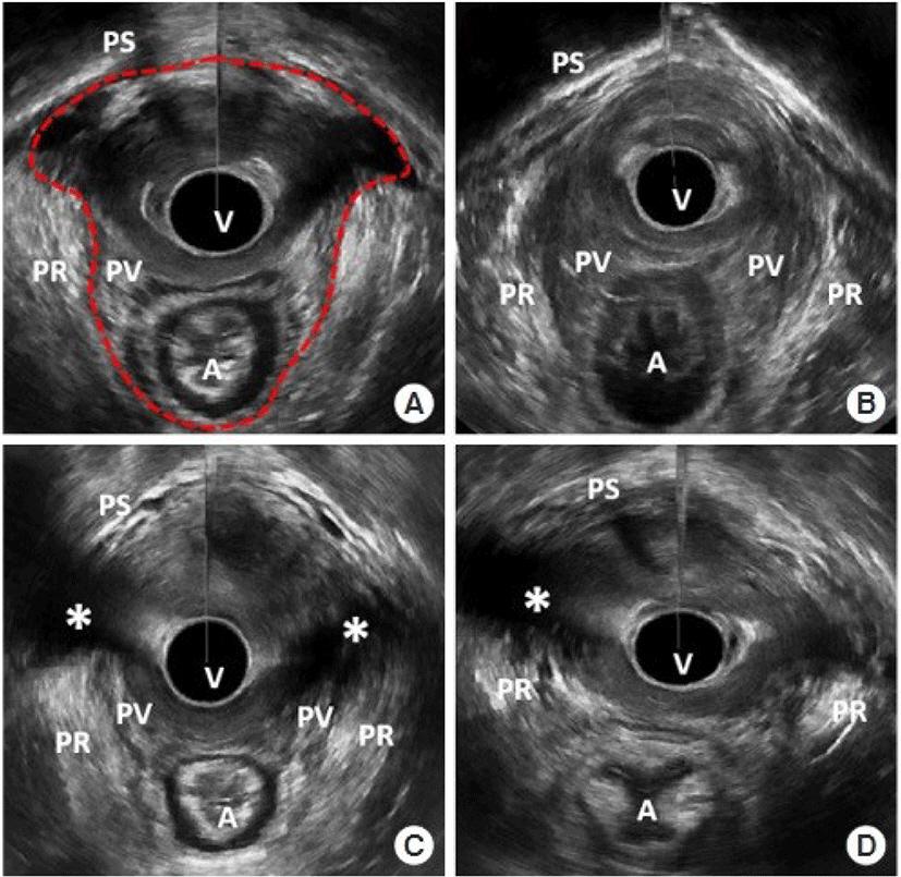
Ultrasound imaging of the anorectum is the most sensitive method for evaluating the
anal complex for the existence of any defect or lesion [30]. Endoanal ultrasound imaging permits the categorization of
lesions according to their whole or partial thickness and degree of disruption. A
common application of anal ultrasonography prior to anus surgery is to identify the
location and extent of anal sphincter problems. Ultrasonography of the 3D pelvic
floor revealed anal sphincter damage and levator ani muscle avulsion in all groups.
In addition, defecography and pelvic MRI are tests that can detect rectoceles and
internal rectal prolapse, as well as see pelvic floor muscle activity during
defecation (Fig. 1) [31,32].
Fig. 1.
3D-pelvic floor ultrasonography. (A) Minimal levator hiatus (MLH), (B)
mild levator ani deficiency (LAD) score, (C) severe LAD score, (D) levator
ani muscle avulsion. Red dot-ted line, MLH area; asterisk, avulsion site. A,
anus; PR, puborectalis muscle; PS, pubic symphysis; PV, pubovisceralis
muscle; V, vaginal [31]. Adapted from
Jeong et al. [31] with
CC-BY-NC.
Anorectal manometry evaluates the resting and squeezed anal sphincter function of
patients [25]. Manometry measures muscular
strength and reservoir function, which includes anorectal sensation, volume
tolerance, and rectal compliance [33]. In the
high-pressure region of the rectum, the recto-anal inhibitory reflex is evaluated
via fast balloon inflation and deflation. A diminished recto-anal inhibitory reflex
is indicative of a neurological disorder. Pudendal nerve terminal motor latency is
used to diagnose pudendal neuropathy or systemic diseases such as diabetes and
chemotherapy. Electrical impulses are administered to the pudendal nerve after an
electrode is affixed to the examiner's finger and directed toward the ischial
spine. The response time at the external anal sphincter level is measured. Normal
reaction time is 2.0±0.2 milliseconds.
Electromyography employs a concentric needle electrode to record the electrical
activity produced by the anal sphincter muscle fibers. Encircling the anal canal,
continuous recordings of the motor units are taken. This test is used to map the
external sphincter and neuromuscular function.
Treatment
FI treatment can be categorized into non-surgical and surgical approaches. We stated
non-medical treatment methods that excluded drug treatment.
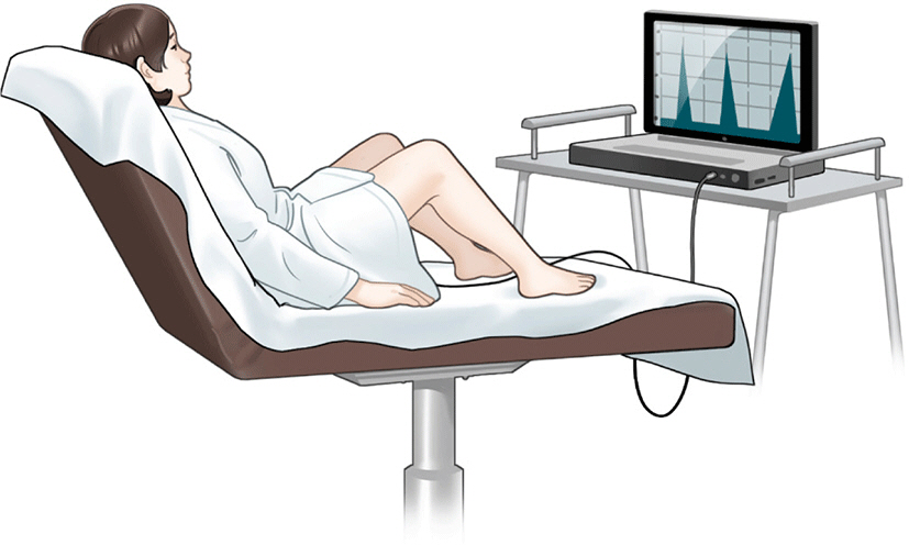
1. Biofeedback therapy
BFT involves the use of electronic devices to monitor physiologic activities,
followed by the transmission of a visual or auditory signal to the patient
(Fig. 2). It makes patients sensitive
to rectal distension and reinforces correct sphincter contraction. Anorectal
manometry or electromyography are utilized to perform the BFT. BFT consists of
rectal sensitivity training, pelvic floor muscle strength training, and
coordination training involving the insertion of an air or water-filled balloon
into the rectum of patients. Patients feel the distension of the balloon and are
instructed to squeeze their pelvic floor muscles to determine the sensation of
rectal filling and prevent leakage [34].
Fig. 2.
Biofeedback therapy. An electrical device capable of monitoring the
physiological activity of the anus is attached to the patient. The
patient looks at the monitor and practices contraction and relaxation of
the anus.
In 2012, a Cochran review of 21 studies involving a total of 1,525 patients did
not find evidence that pelvic floor muscle exercise and BFT were superior to
other treatments. However, it was discovered that BFT was more effective than
pelvic floor muscle exercises alone, such as Kegel, for patients whose
conservative treatments had failed [35].
In a recent randomized controlled trial, patients with FI responded similarly to
oral placebo plus education only, oral placebo plus anorectal manometry-assisted
biofeedback, loperamide plus education only, and loperamide plus anorectal
manometry-assisted biofeedback [36]. BFT
is a noninvasive, first-line treatment option for highly motivated patients
whose medical treatments have failed [37].
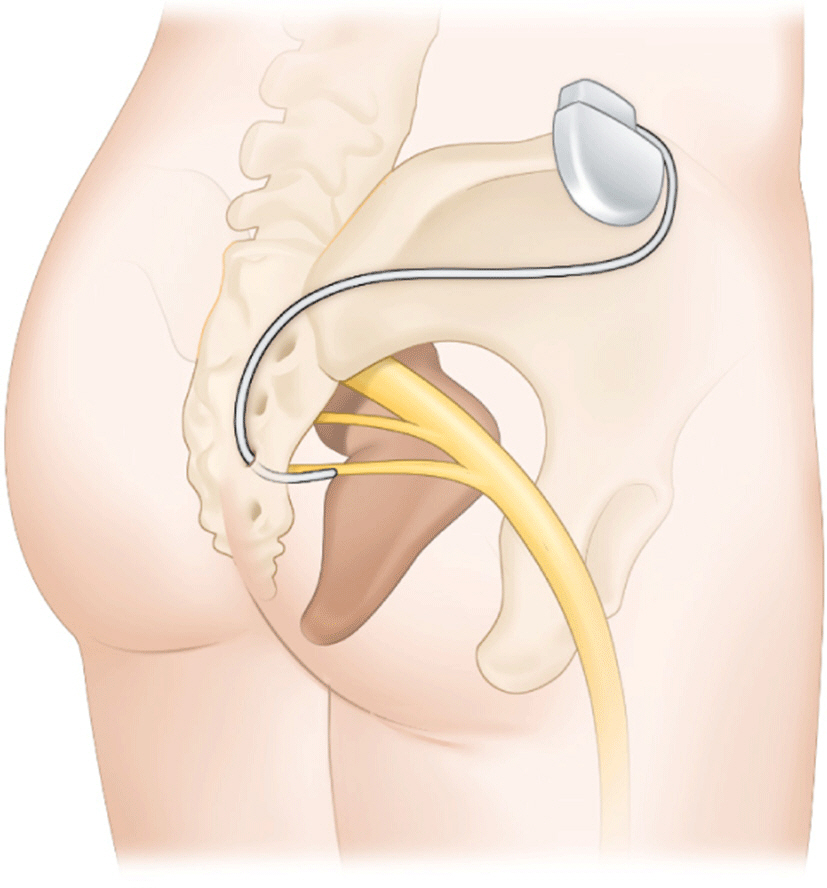
2. Neuromodulation
SNS is a treatment for patients whose conservative treatment has failed. SNS has
been used as a treatment for FI for twenty years, and it is believed to improve
FI. It stimulates sacral nerves and associated muscles by applying a low-voltage
electrical current through the sacral foramen via an implanted electrode. The
surgical placement of a tined lead with electrodes through the S3 sacral foramen
is required. The lead is attached to the pulse generator's battery and
placed beneath the lumbar region of the patient's skin (Fig. 3). Thus, SNS is commonly referred to as
a minimally invasive treatment for FI [38]. Recently, SNS devices that are rechargeable and MRI-safe have
emerged. The estimated battery lifespan of rechargeable devices was 15 years,
whereas the estimated battery lifespan of charge-free devices was 5 to 7 years.
In addition, these new devices offer advantages to patients who require an MRI
[39]. The benefit of SNS is that
there is no incision around the anus. This prevents scarring and infection near
the anus. Numerous studies have been conducted on SNS.
Fig. 3.
Sacral nerve stimulation. An electrical device is inserted in
subcutaneous of patient's lumbar region. This device controls the
anus by providing electrical stimulation to the sacral nerve and its
associated muscles.
The benefit of SNS is that there is no incision around the anus. This prevents
scarring and infection near the anus. According to some reports, SNS is
effective at enhancing FI. In multiple cross-over studies, the incidence of FI
was reduced relative to medical therapy [40].
Percutaneous stimulation of the tibial nerve is an additional nerve modulation
treatment for FI. The tibial nerve and sacral nerve share nerve fibers.
According to some studies, percutaneous tibial nerve stimulation yielded
comparable results to SNS [41].
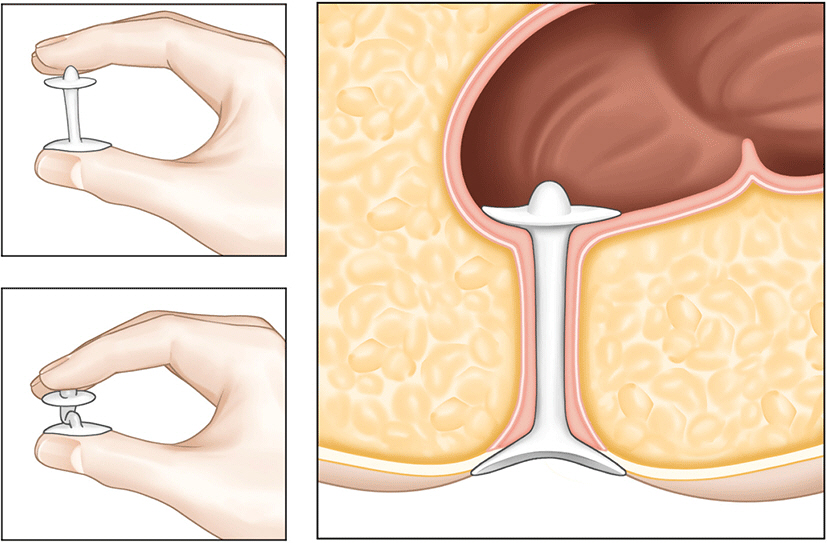
3. Anal implant
Anal plugs are used to prevent stool leakage by occluding the anus. The high
failure rate of anal plugs limits their use as a treatment. However, it is
believed to be beneficial for immobile patients or those residing in nursing
homes with diminished anal-rectal sensation. The Renew Insert anal plug is
designed for self-insertion and natural expulsion with defecation (Fig. 4). Among 91 patients treated with the
Renew insert, 73 (80%) completed all 12 weeks of treatment
("completers"), while 85 (93%) completed at least 1 week of
treatment (modified intention-to-treat cohort). 77% of the 73 participants who
completed the study experienced a 50% reduction in incontinence frequency. 51%
of patients experienced complications, but 98% of these were minor, including
anorectal urgency, irritation, GI discomfort, gas, and hemorrhoids.
Additionally, 78% of participants were pleased with the device [42].
Fig. 4.
Anal plug. This device is designed for self-insertion and natural
expulsion with defecation.
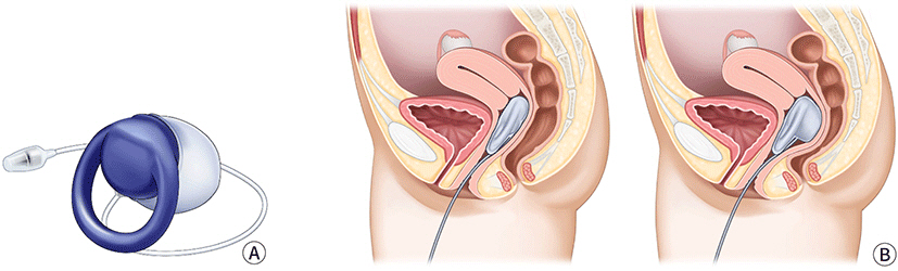
A vaginal bowel control system (Eclipse System) was designed to offer women with
FI a low-risk, reversible treatment (Fig.
5). The system consisted of a silicone-coated base, an inflatable
balloon, and a hand-held pressure control pump. This device is inserted
vaginally. When the balloon is inflated, the vaginal wall is compressed to
prevent stool leakage.
Fig. 5.
Vaginal bowel control system. (A) Vaginal bowel control system. (B)
It is a balloon-shaped structure that is inserted into the vagina. By
applying pressure by hand, the balloon inflates and pressure is applied
to the rectum to control the stool.
Perianal injectables increase the resting anal sphincter tone by acting as a
bulking agent. Its primary use is for the treatment of minor FI caused by
dysfunction of the internal anal sphincter. Bulking materials injected into the
submucosa or inter-sphincteric space increase tissue volume in the high-pressure
zone, resulting in a tighter anus at rest, and can target defective areas of the
internal anal sphincter to create anal canal continuity, if present [34]. The most research has been conducted
on non-animal stabilized hyaluronic acid/dextranomer for the treatment of FI
resistant to conservative therapy. The procedures were performed while the
patient was in the lithotomy, left-lateral, or prone position. Typically, 1 mL
of agent is injected through an endoscope into the deep submucosa at four sites
(typically 3, 6, 9, and 12 o'clock). Active inflammatory bowel disease,
previous anorectal radiation, full-thickness rectal prolapse, and anorectal
malformations are contraindications. Post-injection complications consisted
primarily of transient bleeding, pain, and discomfort that resolved on their own
[43]. Due to concerns regarding the
bulking agent's absorption and migration, additional long-term data are
still required [34].
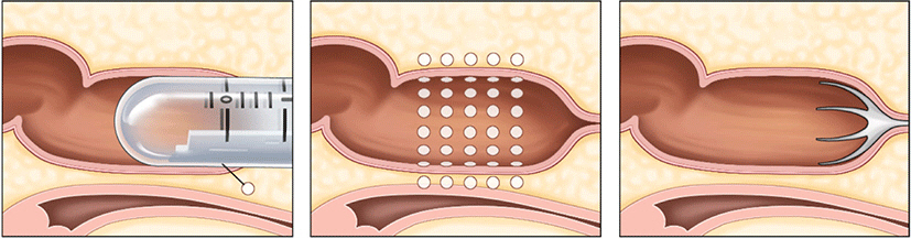
4. SECCA procedure
In 1999, the SECCA® procedure, which involves the application of
temperature-controlled radiofrequency energy to the anal canal, was first used
in Mexico to treat FI.
As a result of the thermal energy delivered, there is an immediate contraction of
collagen fibers, which is followed by permanent shortening via remodeling,
resulting in a tightening of the anal sphincter muscle. In the SECCA®
procedure, radiofrequency is delivered to the anal sphincter while monitoring
temperature and tissue impedance and cooling the probes at the surface to
prevent mucosal damage. If the tissue temperature rises above 85°C at the
electrode tip or 42°C at the anoderm surface, the current is
automatically cut off (Fig. 6).
Fig. 6.
SECCA procedure. A device that uses temperature-controlled
radiofrequency energy is inserted into the anus of the patient. This
device stimulates the patient's anus, causing the anal sphincter
to tighten.
Patients who failed conventional treatments such food modification, medication,
and biofeedback and do not have a clearly visible sphincter dysfunction would be
indication for SECCA® procedure. Typically, the Secca® operation
is carried out as an outpatient while being sedated intravenously and given
local anesthesia. T The patient is in the prone jackknife or lithotomy posture.
Electrodes are positioned at the level of the dentate line and the device is
introduced into the anal canal. The radiofrequency is then transmitted to one
quadrant for 90 seconds. The procedure is then repeated for each of the four
quadrants at a depth of 5 mm. At the site of insertion, complications include
bleeding and ulceration. Occasionally, stitches or surgical therapy may be
necessary [44].
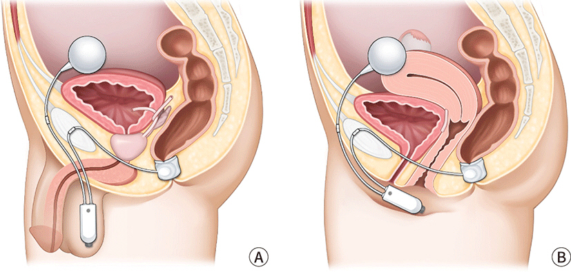
5. Artificial sphincter
Typically, the synthetic sphincter was utilized to treat urine incontinence. The
instrument has been adapted for use around the anus (Fig. 7). To attach an artificial anus to the
patient's body, a transverse perineal incision is typically made to
create a subcutaneous tunnel around the anus. The prosthetic sphincter cuff is
positioned around the anus. The pump is inserted via a pfannensteil incision
that extends to the labia or scrotum. The reservoir is located in the Retzius
space. The gadget provides continence by maintaining a full cuff in a resting
condition. When a patient needs to defecate, fluid is pumped from the cuff to
the reservoir. And the cuff will passively refill. This procedure must require a
sufficient quantity of perineal soft tissue. Because the device is not covered
by thin soft tissue, erosion can occur. It is also necessary to instruct
patients to activate the device independently. Magnetic anal sphincter is a new
artificial anal sphincter option. A single perineal incision is used in the
procedure. The perineal body was dissected to a depth of 5 cm. The left and
right fossa ischiorectalis were then tunneled, with the tip of the coccyx as a
landmark. After the tunneling was completed, the sizing tool was used to
determine the proper sized device. Fluoroscopy was used to confirm the correct
placement and contact of the beads. For the treatment of severe FI patients,
magnetic anal sphincter has shown consistent results [45].
Fig. 7.
Artificial sphincter. A cuff-shaped structure is wrapped around the
anus and inserted (A) male, (B) female. Continence is maintained
normally. Patient needs to press the pump located in the testicles or
perineum for defecation. Then, cuff is closed naturally after
defecation.
Postoperative complications were discovered in a multicenter study [46]. There were 384 device-related
complications in 99 of the 112 patients who received artificial sphincter
treatment. 246 events did not necessitate intervention. However, 73 revisional
operations were performed on 51 (46%) of the patients. 28 (25%) patients
developed infections that necessitated surgical revision, and 41 (37%) patients
had their devices completely removed.
6. Stem cell therapy
In 2008, stem cell therapies were validated. In clinical and experimental
contexts involving hematological, cardiovascular, neurological, digestive,
traumatic, endocrine, renal, and metabolic disorders, stem cell therapy has been
shown to be safe and to have encouraging effects. Hematopoietic stem cells,
mesenchymal stem cells, and adipose-derived stem cells are utilized most
frequently. It has also been demonstrated that stem cell therapy promotes the
healing of acute and subacute anal sphincter injuries. On bioengineered
scaffolds, the formation of an innervated anal sphincter was accomplished by
combining stem cells with normal cells. According to some reports, stem cell
therapy is a successful treatment for FI; nevertheless, additional research in
both animal models and clinical settings is required to determine its efficacy.
Nonetheless, stem cell therapy is one of the potential treatments for FI [47].
7. Surgical option
Surgical therapy is also beneficial for treating FI symptoms. Because surgery is
an intrusive treatment, many practitioners should evaluate the co-morbidities,
socioeconomic circumstances, and activity levels of their patients [48].
Sphincteroplasty aims to restore sphincter architecture and function. Although
anal sphincteroplasty gives temporary symptom relief, it rarely results in
permanent continence recovery. Surgical consequences consist of wound
dehiscence, nerve injury, and infection, and older patients have poorer
outcomes. The most severe side effect of sphincteroplasty is rectovaginal
fistula. For the treatment of obstetric FI, sphincteroplasty has favorable
short- to medium-term outcomes in terms of continence and quality of life [7,49].
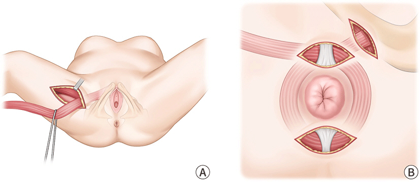
The transposition of the gracilics muscle is an another method that can be used
during surgery (Fig. 8). Patients who did
not respond to conventional medical treatment for FI may be candidates for this
technique. However, there is a considerable risk of problems occurring over the
course of this surgery. Sexual dysfunction and soreness in the area of the
operation site are also common negative outcomes associated with this procedure
[1].
Fig. 8.
Transposition of gracilis muscle. (A) This procedure is mobilization
of the gracilis muscle, (B) transposition of the muscle around the anus
and fixation to the contralateral ischial tuberosity.
Transposition of the antropyloric valve has recently emerged as a therapy for FI.
Beginning the process with a midline laparotomy incision. The colonic hepatic
flexure was mobilized, and the duodenum was kocherized. The gastro epiploic
branches and larger stomach branch curvature were ligated. The left
gastroepiploic pedicle was then utilized to mobilize the antropyloric valve. The
pelvic dissection was followed by rectum mobilization and incision. The portion
of the antropyloric valve was sutured to the proximal rectal end. Invagination
of the graft into the distal rectum. The end of the duodenum was sutured using
perianal skin. Transposition of the antropyloric valve perineally is a unique
procedure for individuals with significantly compromised anorectal function.
Complications of gastrojejunostomy, such as dumping syndrome and alkaline reflux
syndrome, were common with this treatment [50]. In patients who did not respond to the aforementioned treatment
and had a severely diminished quality of life, the creation of an ostomy would
be considered.
Conclusion
With the advent of medicine, patients' lifespans have been lengthened.
Consequently, numerous studies on diseases with a high prevalence among the elderly
have been done. Incontinence of feces is a prevalent condition in the older
population. Patients' quality of life is negatively impacted by this disease,
thus it is evidently a disease that requires control. It has not been demonstrated
which treatment option for FI is the most effective (Table 3). Nevertheless, the majority of therapy techniques often
progress from medicinal treatment to a more intrusive strategy. In addition, when
applied clinically to a patient, multiple treatments are administered concurrently,
as opposed to a single treatment. Examples include SNS, stem cell therapy, and BFT.
It indicates that there is no recognized way for treating FI, but that these various
treatment strategies have a small impact on the improvement of patients'
symptoms. It is believed that research on a definitive therapy for FI will be
required in the foreseeable future. This is important in order to be prepared for an
increase in the number of patients.
Table 3.
Recommendations from professional society guideline of surgical treatment
for fecal incontinence (FI)
Treatment
American college of Gastroenterology
(ACG)
American Society of Colon and Rectal
Surgeons (ASCRS)
Anal sphincter repair
Do not respond to conservative therapy,
have anatomic sphincter defect
Have defect of external anal
sphincter
Redo anal sphincter repair
Not addressed
After failed overlapping sphincteroplasty
should be avoided unless other treatments are not possible
2. Ditah I, Devaki P, Luma HN, Ditah C, Njei B, Jaiyeoba C, et al. Prevalence, trends, and risk factors for fecal incontinence in
United States adults, 2005–2010. Clin Gastroenterol Hepatol 2014;12(4):636-643.
4. Whitehead WE, Borrud L, Goode PS, Meikle S, Mueller ER, Tuteja A, et al. Fecal incontinence in US adults: epidemiology and risk
factors. Gastroenterology 2009;137(2):512-517.
5. Piozzi GN, Kim SH. Robotic intersphincteric resection for low rectal cancer:
technical controversies and a systematic review on the perioperative,
oncological, and functional outcomes. Ann Coloproctol 2021;37(6):351-367.
6. Eldamshety O, Kotb S, Khater A, Roshdy S, Elnahas W, Zahi MS, et al. Early and late functional outcomes of anal sphincter-sparing
procedures with total mesorectal excision for anorectal
adenocarcinoma. Ann Coloproctol 2020;36(3):148-154.
7. Muñoz-Duyos A, Lagares-Tena L, Ribas Y, Baanante JC, Navarro-Luna A. Critical appraisal of international guidelines for the management
of fecal incontinence in adults: is it possible to define what to do in
different clinical scenarios? Tech Coloproctol 2022;26(1):1-17.
9. De Ligny WR, Kerkhof MH, Ruiz-Zapata AM. Regenerative medicine as a therapeutic option for fecal
incontinence: a systematic review of preclinical and clinical
studies. Am J Obstet Gynecol 2019;220(2):142-154.
11. Simren M, Palsson OS, Whitehead WE. Update on Rome IV criteria for colorectal disorders: implications
for clinical practice. Curr Gastroenterol Rep 2017;19(4):15
12. Whitehead WE, Simren M, Busby-Whitehead J, Heymen S, van Tilburg MAL, Sperber AD, et al. Fecal incontinence diagnosed by the Rome IV criteria in the
United States, Canada, and the United Kingdom. Clin Gastroenterol Hepatol 2020;18(2):385-391.
17. Pinsk I, Czeiger D, Lichtman D, Reshef A. The long-term effect of standardized anal dilatation for chronic
anal fissure on anal continence. Ann Coloproctol 2021;37(2):115-119.
18. Egal A, Etienney I, Atienza P. Endorectal advancement flap with muscular plication in anovaginal
and anterior perineal fistulas. Ann Coloproctol 2021;37(3):141-145.
19. Lee KH, Hyun K, Yoon SG, Lee JK. Minimal lateral internal sphincterotomy (LIS): is it enough to
cut less than the conventional tailored LIS? Ann Coloproctol 2021;37(5):275-280.
20. Lopez MPJ, Onglao MAS, Monroy Iii HJ. Initial experience with video-assisted anal fistula treatment in
the Philippines. Ann Coloproctol 2020;36(2):112-118.
22. Markland AD, Dunivan GC, Vaughan CP, Rogers RG. Anal intercourse and fecal incontinence: evidence from the
2009–2010 national health and nutrition examination
survey. Am J Gastroenterol 2016;111(2):269-274.
25. Paquette IM, Varma MG, Kaiser AM, Steele SR, Rafferty JF. The American Society of Colon and Rectal Surgeons’
clinical practice guideline for the treatment of fecal
incontinence. Dis Colon Rectum 2015;58(7):623-636.
29. Rockwood TH, Church JM, Fleshman JW, Kane RL, Mavrantonis C, Thorson AG, et al. Fecal incontinence quality of life scale: quality of life
instrument for patients with fecal incontinence. Dis Colon Rectum 2000;43(1):9-16.
31. Jeong HY, Yang SJ, Cho DH, Park DH, Lee JK. Comparison of 3-dimensional pelvic floor ultrasonography and
defecography for assessment of posterior pelvic floor
disorders. Ann Coloproctol 2020;36(4):256-263.
32. Yune Y, Jeong HY, Park DH, Lee JK. Three-dimensional pelvic floor ultrasound assessment of pelvic
organ prolapse: minimal levator hiatus and levator ani deficiency
score. Ann Coloproctol 2021;37(5):291-297.
33. Fitzpatrick M, O’brien C, O’connell PR, O’herlihy C. Patterns of abnormal pudendal nerve function that are associated
with postpartum fecal incontinence. Am J Obstet Gynecol 2003;189(3):730-735.
36. Eric Jelovsek J, Markland AD, Whitehead WE, Barber MD, Newman DK, Rogers RG, et al. Controlling faecal incontinence in women by performing anal
exercises with biofeedback or loperamide: a randomised clinical
trial. Lancet Gastroenterol Hepatol 2019;4(9):698-710.
39. De Wachter S, Knowles CH, Elterman DS, Kennelly MJ, Lehur PA, Matzel KE, et al. New technologies and applications in sacral neuromodulation: an
update. Adv Ther 2020;37:637-643.
41. Knowles CH, Horrocks EJ, Bremner SA, Stevens N, Norton C, O’Connell PR, et al. Percutaneous tibial nerve stimulation versus sham electrical
stimulation for the treatment of faecal incontinence in adults (CONFIDeNT):
a double-blind, multicentre, pragmatic, parallel-group, randomised
controlled trial. Lancet 2015;386(10004):1640-1648.
42. Lukacz ES, Segall MM, Wexner SD. Evaluation of an anal insert device for the conservative
management of fecal incontinence. Dis Colon Rectum 2015;58(9):892-898.
43. Maeda Y, Laurberg S, Norton C. Perianal injectable bulking agents as treatment for faecal
incontinence in adults. Cochrane Database Syst Rev 2013;(2):CD007959.
44. Frascio M, Mandolfino F, Imperatore M, Stabilini C, Fornaro R, Gianetta E, et al. The SECCA procedure for faecal incontinence: a
review. Colorectal Dis 2013;16(3):167-172.
45. Pakravan F, Helmes C. Magnetic anal sphincter augmentation in patients with severe
fecal incontinence. Dis Colon Rectum 2015;58(1):109-114.
46. Wong WD, Congliosi SM, Spencer MP, Corman ML, Tan P, Opelka FG, et al. The safety and efficacy of the artificial bowel sphincter for
fecal incontinence. Dis Colon Rectum 2002;45(9):1139-1153.
47. Trébol J, Carabias-Orgaz A, García-Arranz M, García-Olmo D. Stem cell therapy for faecal incontinence: current state and
future perspectives. World J Stem Cells 2018;10(7):82-105.
48. Forte ML, Andrade KE, Lowry AC, Butler M, Bliss DZ, Kane RL. Systematic review of surgical treatments for fecal
incontinence. Dis Colon Rectum 2016;59(5):443-469.
49. Pla-Martí V, Martín-Arévalo J, Martí-Fernández R, Moro-Valdezate D, García-Botello S, Espí-Macías A, et al. Long-term evolution of continence and quality of life after
sphincteroplasty for obstetric fecal incontinence. Ann Coloproctol 2022;38(1):13-19.
Is It a Refractory Disease?- Fecal Incontinence; beyond
Medication
Fig. 1.
3D-pelvic floor ultrasonography. (A) Minimal levator hiatus (MLH), (B)
mild levator ani deficiency (LAD) score, (C) severe LAD score, (D) levator
ani muscle avulsion. Red dot-ted line, MLH area; asterisk, avulsion site. A,
anus; PR, puborectalis muscle; PS, pubic symphysis; PV, pubovisceralis
muscle; V, vaginal [31]. Adapted from
Jeong et al. [31] with
CC-BY-NC.
Fig. 2.
Biofeedback therapy. An electrical device capable of monitoring the
physiological activity of the anus is attached to the patient. The
patient looks at the monitor and practices contraction and relaxation of
the anus.
Fig. 3.
Sacral nerve stimulation. An electrical device is inserted in
subcutaneous of patient's lumbar region. This device controls the
anus by providing electrical stimulation to the sacral nerve and its
associated muscles.
Fig. 4.
Anal plug. This device is designed for self-insertion and natural
expulsion with defecation.
Fig. 5.
Vaginal bowel control system. (A) Vaginal bowel control system. (B)
It is a balloon-shaped structure that is inserted into the vagina. By
applying pressure by hand, the balloon inflates and pressure is applied
to the rectum to control the stool.
Fig. 6.
SECCA procedure. A device that uses temperature-controlled
radiofrequency energy is inserted into the anus of the patient. This
device stimulates the patient's anus, causing the anal sphincter
to tighten.
Fig. 7.
Artificial sphincter. A cuff-shaped structure is wrapped around the
anus and inserted (A) male, (B) female. Continence is maintained
normally. Patient needs to press the pump located in the testicles or
perineum for defecation. Then, cuff is closed naturally after
defecation.
Fig. 8.
Transposition of gracilis muscle. (A) This procedure is mobilization
of the gracilis muscle, (B) transposition of the muscle around the anus
and fixation to the contralateral ischial tuberosity.
Fig. 1.
Fig. 2.
Fig. 3.
Fig. 4.
Fig. 5.
Fig. 6.
Fig. 7.
Fig. 8.
Is It a Refractory Disease?- Fecal Incontinence; beyond
Medication
Risk factors of fecal incontinence
Category
Factors
Patient
Aging, obesity, smoking, post-menopause,
nursing home residency
Lack of ability to defer
defecation for 15 minutes
0
4
Score of 0=perfect continence;
24=totally incontinence never, 0; rarely, 1/ month;
sometimes, >1/ month but <1 a week; weekly, ≥1/
week but <1 a day; daily, ≥1/ day
Fecal incontinence quality of
life (FIQL) scale composition
Questions: scale
1-lifestyle
I cannot do many of
things I want to do. I am afraid to go
out. It is important to plan my schedule
around my bowel pattern. I cut down on how
much I eat before I go out. It is difficult
for me to get out and do things like going to a movie or to
church. I avoid traveling by plane or
train. I avoid
traveling. I avoid visiting
friends. I avoid going out to
eat. I avoid staying overnight away from
home.
Scale 2. coping/behavior
I have sex less often
than I would like to. The possibility of bowel
accidents is always on my mind. I feel I have
no control over my bowels. Whenever I go
someplace new, I specifically locate where the bathrooms
are. I worry about not being able to get to
the toilet in time. I worry about bowel
accidents. I try to prevent bowel accidents by
staying very near a bathroom. I can’t
hold my bowel movement long enough to get to the
bathroom. Whenever I am away from home, I try
to stay near a restroom as much as possible.
Question: scale
3-depression/self perception
In general, would you
say your health is excellent, very good, good, fair, and
poor. I am afraid to have
sex. I feel different from other
people. I enjoy life
less. I feel like I am not a healthy
person. I feel
depressed. During past month, have you felt so
sad, discouraged, hopeless, or had so many problems that you
wondered if anything was worthwhile.
Question: scale
4-embarrassment
I leak stool without
even knowing it. I worry about others smelling
stool on me. I feel ashamed.
Recommendations from professional society guideline of surgical treatment
for fecal incontinence (FI)
Treatment
American college of Gastroenterology
(ACG)
American Society of Colon and Rectal
Surgeons (ASCRS)
Anal sphincter repair
Do not respond to conservative therapy,
have anatomic sphincter defect
Have defect of external anal
sphincter
Redo anal sphincter repair
Not addressed
After failed overlapping sphincteroplasty
should be avoided unless other treatments are not possible