1Shoulder & Elbow Clinic, Department of Orthopaedic Surgery, College of Medicine, Kyung Hee University Hospital, Seoul, Korea
*Corresponding author: Sung Min Rhee,
Shoulder & Elbow Clinic, Department of Orthopaedic Surgery, College of
Medicine, Kyung Hee University Hospital, 23, Kyungheedae-ro, Dongdaemun-gu,
Seoul 02447, Korea, E-mail: minrhee77@gmail.com
• Received: November 17, 2024 • Revised: December 24, 2024 • Accepted: January 7, 2025
This is an Open-Access article distributed under the terms of the
Creative Commons Attribution Non-Commercial License (http://creativecommons.org/licenses/by-nc/4.0) which permits
unrestricted non-commercial use, distribution, and reproduction in any
medium, provided the original work is properly cited.
Shoulder diseases pose a significant health challenge for older adults, often
causing pain, functional decline, and decreased independence. This narrative
review explores how deep learning (DL) can address diagnostic challenges by
automating tasks such as image segmentation, disease detection, and motion
analysis. Recent research highlights the effectiveness of DL-based convolutional
neural networks and machine learning frameworks in diagnosing various shoulder
pathologies. Automated image analysis facilitates the accurate assessment of
rotator cuff tear size, muscle degeneration, and fatty infiltration in MRI or CT
scans, frequently matching or surpassing the accuracy of human experts.
Convolutional neural network-based systems are also adept at classifying
fractures and joint conditions, enabling the rapid identification of common
causes of shoulder pain from plain radiographs. Furthermore, advanced techniques
like pose estimation provide precise measurements of the shoulder joint's
range of motion and support personalized rehabilitation plans. These automated
approaches have also been successful in quantifying local osteoporosis,
utilizing machine learning-derived indices to classify bone density status. DL
has demonstrated significant potential to improve diagnostic accuracy,
efficiency, and consistency in the management of shoulder diseases in older
patients. Machine learning-based assessments of imaging data and motion
parameters can help clinicians optimize treatment plans and improve patient
outcomes. However, to ensure their generalizability, reproducibility, and
effective integration into routine clinical workflows, large-scale, prospective
validation studies are necessary. As data availability and computational
resources increase, the ongoing development of DL-driven applications is
expected to further advance and personalize musculoskeletal care, benefiting
both healthcare providers and the aging population.
Shoulder diseases pose a significant health burden on the aging population,
affecting millions of individuals worldwide [1–3]. Common conditions
such as rotator cuff tears, impingement syndrome, osteoarthritis, and adhesive
capsulitis not only cause pain but also significantly impair the daily lives of
patients by restricting their mobility and independence [1,4–8]. Timely and accurate diagnosis of these
conditions is crucial for optimizing treatment outcomes and enhancing patient
quality of life. However, traditional diagnostic tools, such as X-rays, MRI, and
ultrasound, face challenges including variability in interpretation and limited
availability in resource-constrained environments [9]. Furthermore, these methods struggle to accurately and
objectively measure joint range of motion, which further compromises their
effectiveness in diagnosing musculoskeletal conditions [10].
Recent advances in artificial intelligence (AI), especially in the area of deep
learning (DL), have revolutionized the diagnosis of shoulder diseases [11–14]. DL algorithms leverage artificial neural networks, modeled
after the human brain, to process and analyze vast amounts of data with
exceptional accuracy [15]. These
algorithms can detect subtle patterns in medical images that may be overlooked
by even experienced radiologists. They also analyze complex movements and
postures through pose estimation techniques. By minimizing diagnostic errors,
improving consistency, and facilitating detailed motion analysis, DL algorithms
are widely applicable in imaging and movement assessment, transforming sectors
like healthcare, rehabilitation, and biomechanics.
Objectives
This paper aims to explore recent studies on the application of DL in diagnosing
shoulder diseases in older adults.
Ethics statement
As this study is a literature review, it did not require institutional review board
approval or individual consent.
The analysis of rotator cuff muscles/tendons and fatty infiltrations using
artificial intelligence
In 2020, Taghizadeh et al. introduced an AI model specifically designed to
automatically assess rotator cuff muscle degeneration by analyzing both atrophy and
fatty infiltration in CT images [14]. This
model utilized a convolutional neural network (CNN) to automatically evaluate
degeneration, including atrophy and fatty infiltration, in preoperative shoulder CT
scans of patients with glenohumeral osteoarthritis. The CNN was tested on
retrospective data from 103 CT scans and achieved Dice similarity coefficients that
were comparable to those of manual radiologist segmentations. It demonstrated high
accuracy in measuring atrophy (R2=0.87), fatty infiltration
(R2=0.91), and overall degeneration (R2=0.91). These
findings highlight the potential of DL to provide efficient and reliable evaluations
of rotator cuff muscles preoperatively.
Similarly, Ro et al. developed a DL framework that utilizes MRI to evaluate factors
such as the occupation ratio and fatty infiltration in the supraspinatus muscle of
patients with rotator cuff tears [12]. This
study employed a deep-learning framework to analyze the occupation ratio and fatty
infiltration in the supraspinatus muscle using shoulder MRI. A full CNN facilitated
rapid and precise segmentation of the supraspinatus muscle and fossa, achieving high
Dice similarity coefficients (0.97 for the fossa and 0.94 for the muscle) along with
excellent sensitivity and specificity. Fatty infiltration was quantified using a
region-based Otsu thresholding method, which revealed significant differences across
Goutallier grades (P<0.0001) [16] and
demonstrated a moderate negative correlation with the occupation ratio
(ρ=−0.75, P<0.0001) [17]. These findings indicate that integrating DL with automated thresholding
techniques offers an objective and efficient means of quantifying key indices in
shoulder MRI, thereby enhancing diagnostic accuracy and consistency.
Detection of shoulder pathologies including rotator cuff tears and
fractures
Recently, DL technology has been employed to automate the segmentation and detection
of rotator cuff tears using MRI.
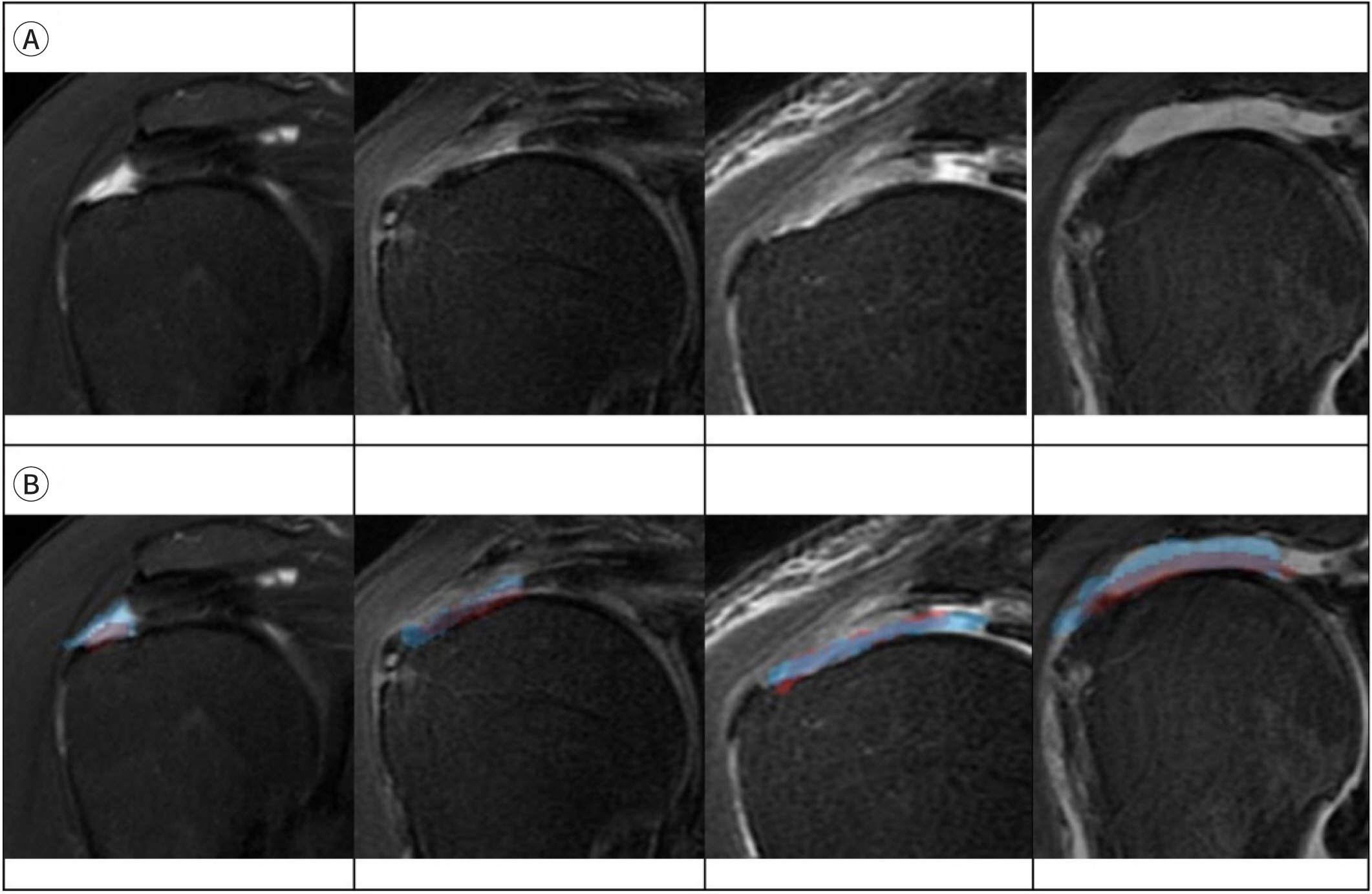
Lee et al. developed a DL model utilizing a 3D U-Net CNN to detect, segment, and
visualize rotator cuff tear lesions in three dimensions using MRI data from 303
patients [18]. The model, trained and
validated on labeled MRI datasets, demonstrated robust performance. It achieved a
Dice coefficient of 94.3%, a sensitivity of 97.1%, a specificity of 95.0%, a
precision of 84.9%, an F1-score of 90.5%, and a Youden index of 91.8% (Fig. 1).
Fig. 1.
Segmentation results corresponding to the rotator cuff tear site. (A)
Original MRI images displaying the presence of a rotator cuff tear. (B) The
red region represents the area manually labeled by shoulder specialists,
while the blue region indicates the area segmented by the proposed deep
learning model. Adapted from Lee et al. [18] with CC-BY.
Hashimoto et al. assessed the diagnostic capabilities of a CNN in detecting and
classifying rotator cuff tears, using 1,169 anteroposterior shoulder radiographs.
These were categorized into four groups: intact, small, medium, and large-to-massive
tears [19]. In binary classification tasks,
the CNN achieved a sensitivity of 92%, a specificity of 69%, an accuracy of 86%, and
an area under the receiver operating curve (AUC) of 0.88. The CNN outperformed
orthopedic surgeons in both detection and classification accuracy, demonstrating its
potential as a reliable tool for diagnosing rotator cuff tears from plain
radiographs.
A recent meta-analysis demonstrated that AI could perform comparably to clinicians in
detecting fractures, highlighting its potential for broader applications in
orthopedics. Magnéli et al. developed and evaluated a CNN for classifying
fractures in shoulder radiographs, focusing on proximal humeral fractures (PHF)
based on the AO/OTA classification system, with secondary objectives for diaphyseal
humerus, clavicle, and scapula fractures [20]. The CNN, trained on a dataset of 6,172 examinations, achieved an
overall AUC of 0.89 for fracture classification. Notably, the AUC for PHF classes
exceeded 0.90. The model also demonstrated excellent AUCs for diaphyseal humerus
(0.97) and clavicle fractures (0.96), and a good performance for scapula fractures
(0.87). Furthermore, Grauhan et al. developed a model capable of identifying a
variety of common causes of shoulder pain on radiographs, extending beyond fractures
to include conditions such as PHF, dislocations, periarticular calcifications,
osteoarthritis, osteosynthesis, and joint prostheses [11]. This study utilized the ResNet-50 architecture to detect
common causes of shoulder pain—such as fractures, dislocations,
osteoarthritis, periarticular calcifications, osteosynthesis, and
endoprosthesis—from plain radiographs. Trained on 2,700 radiographs and
evaluated on a separate annotated dataset, the model demonstrated high accuracy. The
CNN achieved excellent performance, with AUC values of 0.871 for fractures, 0.896
for joint dislocations, 0.945 for osteoarthritis, and 0.800 for periarticular
calcifications. It also detected osteosynthesis and endoprosthesis with high
accuracy, achieving AUC values of 0.998 and 1.0, respectively. Sensitivity and
specificity varied by condition, with values of 0.75 and 0.86 for fractures, 0.95
and 0.65 for joint dislocations, 0.90 and 0.86 for osteoarthritis, and 0.60 and 0.89
for calcifications. These results underscore the potential of CNNs to aid clinicians
by prioritizing worklists and improving diagnostic efficiency in high-workload
settings.
Detection of local osteoporosis in the proximal humerus
Li et al. developed a diagnostic method using machine learning to assess local
osteoporosis in the proximal humerus by analyzing demographic data, bone density,
and X-ray ratios [21]. The study involved a
cohort of 97 patients (76 females and 21 males with an average age of 73 years),
categorized into groups based on bone density: normal (25 patients), osteopenia (35
patients), and osteoporosis (37 patients). Utilizing the modified Tingart index
[22], a decision tree was employed to
identify critical diagnostic indicators, including the humeral shaft medullary
cavity ratio (M2/M4), age, and sex. An M2/M4 ratio below 1.13 was indicative of
local osteoporosis, whereas a ratio of 1.13 or higher, when analyzed alongside age
and sex, helped differentiate between osteoporosis, osteopenia, and normal bone
density. The decision tree achieved accuracies of 76.27% in the training set and
78.95% in the validation set. Additionally, multinomial logistic regression
validated significant associations of M2/M4, age, and sex with osteoporosis.
Analysis of shoulder range of motion using machine learning
Measuring shoulder joint angles accurately has been challenging due to the complexity
of shoulder motion and its intricate rotational axes. Recently, pose estimation, a
computer vision technique that utilizes machine learning, has garnered significant
attention [23,24]. This technology predicts the positions and orientations of human
joints or key points from images or videos, enabling detailed analysis of movements
and postures [25]. In a recent study, the
integration of pose estimation AI with machine learning has demonstrated a promising
approach to estimating the range of motion of the shoulder with remarkable
precision, paving the way for advancements in sports biomechanics and rehabilitation
(Fig. 2).
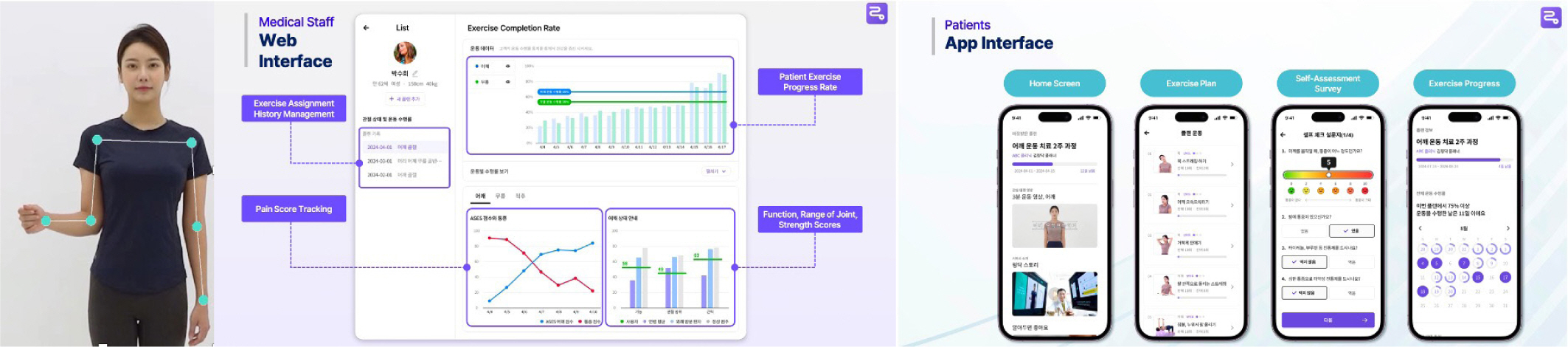
Fig. 2.
A company utilizes machine learning-based pose estimation technology to
measure a patient's range of motion, analyze the patient's
current condition based on the results, and assign the most suitable
rehabilitation exercises. This figure is used with permission from Itphy,
Inc.
Takigami et al. employed pose estimation AI in conjunction with a machine learning
model to estimate the internal and external rotation angles of the shoulder [26]. They processed videos of 10 healthy male
volunteers (average age 37.7 years) into 10,608 images to develop parameters for
training the model. Using smartphone angle measurements as the ground truth, the AI
model demonstrated a correlation coefficient of 0.971 and a mean absolute error of
5.778 using linear regression. With Light GBM, it achieved a correlation coefficient
of 0.999 and an mean absolute error of 0.945. This method offers a precise and
efficient way to measure shoulder rotation angles, showing great potential for
applications in sports biomechanics and rehabilitation.
Ramkumar et al. validated a motion-based machine learning software development kit
designed to assess shoulder range of motion. They compared its accuracy with that of
manual goniometer measurements across four motion arcs: abduction, forward flexion,
internal rotation, and external rotation [27]. Utilizing a mobile application, 10 subjects each performed the motions
five times. The software development kit recorded mean angular differences of less
than 5° for all motions (P>0.05), with specific mean differences of
–3.7° for abduction, –4.9° for forward flexion,
–2.4° for internal rotation, and –2.6° for external
rotation.
Conclusion
The use of DL in diagnosing shoulder diseases among older patients has shown
considerable promise in several areas. These include analyzing rotator cuff muscle
degeneration, detecting pathologies such as rotator cuff tears and fractures,
evaluating local osteoporosis in the proximal humerus, and accurately measuring the
shoulder's range of motion. DL models, which employ sophisticated
architectures like CNNs and incorporate machine learning algorithms, consistently
achieve high levels of accuracy, sensitivity, and specificity in medical imaging
tasks. These models often outperform traditional diagnostic techniques and expert
clinicians.
Authors' contributions
All work was done by Sung Min Rhee.
Conflict of interest
Sung Min Rhee is the Chief Executive Officer of Itphy Inc., the company that
provided Fig. 2. Otherwise, there are no
conflicts of interest to declare.
Funding
Not applicable.
Data availability
Not applicable.
Acknowledgments
Not applicable.
Supplementary materials
Not applicable.
References
1. Gumina S, Kim H, Jung Y, Song HS. Rotator cuff degeneration and healing after rotator cuff
repair. Clin Shoulder Elb 2023;26(3):323-329.
2. Lucas J, van Doorn P, Hegedus E, Lewis J, van der Windt D. A systematic review of the global prevalence and incidence of
shoulder pain. BMC Musculoskelet Disord 2022;23(1):1073
3. Hodgetts CJ, Leboeuf-Yde C, Beynon A, Walker BF. Shoulder pain prevalence by age and within occupational groups: a
systematic review. Arch Physiother 2021;11(1):24
4. Kim YT, Kim TY, Lee JB, Hwang JT. Glenohumeral versus subacromial steroid injections for
impingement syndrome with mild stiffness: a randomized controlled
trial. Clin Shoulder Elb 2023;26(4):390-396.
5. Mardani-Kivi M, Hashemi-Motlagh K, Darabipour Z. Arthroscopic release in adhesive capsulitis of the shoulder: a
retrospective study with 2 to 6 years of follow-up. Clin Shoulder Elb 2021;24(3):172-177.
6. Hones KM, Hao KA, Buchanan TR, Trammell AP, Wright JO, Wright TW, et al. Does preoperative forward elevation weakness affect clinical
outcomes in anatomic or reverse total shoulder arthroplasty patients with
glenohumeral osteoarthritis and intact rotator cuff? Clin Shoulder Elb 2024;27(3):316-326.
7. Rhee SM, Youn SM, Kim CH, Chang GW, Kim SY, Ham HJ, et al. Rotator cuff repairs with all-suture tape anchors: no difference
in outcomes between with or without all-suture tape anchors. Knee Surg Sports Traumatol Arthrosc 2023;31(9):4060-4067.
8. Ko YW, Park JH, Youn SM, Rhee YG, Rhee SM. Effects of comorbidities on the outcomes of manipulation under
anesthesia for primary stiff shoulder. J Shoulder Elbow Surg 2021;30(8):E482-E492.
9. Malavolta EA, Assunção JH, Gracitelli MEC, Yen TK, Bordalo-Rodrigues M, Ferreira Neto AA. Accuracy of magnetic resonance imaging (MRI) for subscapularis
tear: a systematic review and meta-analysis of diagnostic
studies. Arch Orthop Trauma Surg 2019;139(5):659-667.
10. Elrahim RMA, Embaby EA, Ali MF, Kamel RM. Inter-rater and intra-rater reliability of Kinovea software for
measurement of shoulder range of motion. Bull Fac Phys Ther 2016;21(2):80-87.
11. Grauhan NF, Niehues SM, Gaudin RA, Keller S, Vahldiek JL, Adams LC, et al. Deep learning for accurately recognizing common causes of
shoulder pain on radiographs. Skeletal Radiol 2022;51(2):355-362.
12. Ro K, Kim JY, Park H, Cho BH, Kim IY, Shim SB, et al. Deep-learning framework and computer assisted fatty infiltration
analysis for the supraspinatus muscle in MRI. Sci Rep 2021;11(1):15065
13. Shariatnia MM, Ramazanian T, Sanchez-Sotelo J, Maradit Kremers H. Deep learning model for measurement of shoulder critical angle
and acromion index on shoulder radiographs. JSES Rev Rep Tech 2022;2(3):297-301.
14. Taghizadeh E, Truffer O, Becce F, Eminian S, Gidoin S, Terrier A, et al. Deep learning for the rapid automatic quantification and
characterization of rotator cuff muscle degeneration from shoulder CT
datasets. Eur Radiol 2021;31(1):181-190.
15. Alzubaidi L, Al-Dulaimi K, Salhi A, Alammar Z, Fadhel MA, Albahri AS, et al. Comprehensive review of deep learning in orthopaedics:
applications, challenges, trustworthiness, and fusion. Artif Intell Med 2024;155:102935
18. Lee SH, Lee J, Oh KS, Yoon JP, Seo A, Jeong Y, et al. Automated 3-dimensional MRI segmentation for the posterosuperior
rotator cuff tear lesion using deep learning algorithm. PLoS One 2023;18(5):e0284111.
19. Hashimoto E, Maki S, Ochiai N, Ise S, Inagaki K, Hiraoka Y, et al. Automated detection and classification of the rotator cuff tear
on plain shoulder radiograph using deep learning. J Shoulder Elbow Surg 2024;33(8):1733-1739.
20. Magnéli M, Ling P, Gislén J, Fagrell J, Demir Y, Arverud ED, et al. Deep learning classification of shoulder fractures on plain
radiographs of the humerus, scapula and clavicle. PLoS One 2023;18(8):e0289808.
21. Li G, Wu N, Zhang J, Song Y, Ye T, Zhang Y, et al. Proximal humeral bone density assessment and prediction analysis
using machine learning techniques: An innovative approach in medical
research. Heliyon 2024;10(15):e35451.
22. Tingart MJ, Zurakowski D, Warner JJP, Apreleva M, von Stechow D. The cortical thickness of the proximal humeral diaphysis predicts
bone mineral density of the proximal humerus. J Bone Joint Surg Br 2003;85(4):611-617.
23. van den Hoorn W, Lavaill M, Cutbush K, Gupta A, Kerr G. Comparison of shoulder range of motion quantified with mobile
phone video-based skeletal tracking and 3D motion capture: preliminary
study. Sensors 2024;24(2):534
25. Sun K, Xiao B, Liu D, Wang J. Deep high-tesolution representation learning for human pose estimation.
Proceedings of the 2019 IEEE/CVF Conference on Computer Vision and Pattern
Recognition (CVPR). 2019 Jun 15-20; Long Beach, CA. Piscataway (NJ): IEEE; 2019. p. p. 5693-5703.
26. Takigami S, Inui A, Mifune Y, Nishimoto H, Yamaura K, Kato T, et al. Estimation of shoulder joint rotation angle using tablet device
and pose estimation artificial intelligence model. Sensors 2024;24(9):2912
27. Ramkumar PN, Haeberle HS, Navarro SM, Sultan AA, Mont MA, Ricchetti ET, et al. Mobile technology and telemedicine for shoulder range of motion:
validation of a motion-based machine-learning software development
kit. J Shoulder Elbow Surg 2018;27(7):1198-1204.
Diagnosis of SLAP lesions on shoulder MRI using a 2.5D deep learning and ensemble learning framework Hongyu Wang, Qingyun Xue, Lei Shi, Fei Wang, Guanghan Gao, Lin Wang Frontiers in Surgery.2026;[Epub] CrossRef
Application of deep learning for diagnosis of shoulder diseases in
older adults: a narrative review
Fig. 1.
Segmentation results corresponding to the rotator cuff tear site. (A)
Original MRI images displaying the presence of a rotator cuff tear. (B) The
red region represents the area manually labeled by shoulder specialists,
while the blue region indicates the area segmented by the proposed deep
learning model. Adapted from Lee et al. [18] with CC-BY.
Fig. 2.
A company utilizes machine learning-based pose estimation technology to
measure a patient's range of motion, analyze the patient's
current condition based on the results, and assign the most suitable
rehabilitation exercises. This figure is used with permission from Itphy,
Inc.
Fig. 1.
Fig. 2.
Application of deep learning for diagnosis of shoulder diseases in
older adults: a narrative review