1Department of Anesthesiology and Pain Medicine, Ewha Womans University Mokdong Hospital, Ewha Womans University College of Medicine, Seoul, Korea
*Corresponding author: Won-joong Kim,
Department of Anesthesiology and Pain Medicine, Ewha Womans University Mokdong
Hospital, Ewha Womans University College of Medicine, 1071 Anyangcheon-ro,
Yangcheon-gu, Seoul 07985, Korea E-mail:
ickypoo@ewha.ac.kr
• Received: March 13, 2024 • Revised: April 15, 2024 • Accepted: April 16, 2024
This is an Open-Access article distributed under the terms of the
Creative Commons Attribution Non-Commercial License (http://creativecommons.org/licenses/by-nc/4.0) which permits
unrestricted non-commercial use, distribution, and reproduction in any
medium, provided the original work is properly cited.
Although sciatica is commonly associated with lumbar spinal issues, it is
important to acknowledge that non-spinal factors can also play a significant
role in this condition. This is particularly relevant for female patients, in
whom gynecologic conditions can lead to secondary sciatic neuropathy. Herein, we
report the case of a 66-year-old woman who experienced posterolateral right
lower extremity radiating pain. We initially performed a lumbar transforaminal
epidural steroid injection, but the pain persisted. Subsequently, hip MRI
revealed sciatic neuropathy adjacent to the pedunculated portions of a uterine
myoma. We then performed a sub-gluteal sciatic nerve block under ultrasound
guidance, resulting in significant relief of her pain. In conclusion, hip MRI
can be helpful for the differential diagnosis of sciatica, and ultrasound-guided
sciatic nerve block can be considered an appropriate and effective treatment
option.
Sciatica refers to pain that travels downward from the buttocks along the path of the
sciatic nerve, originating from the L4 through S2 nerve roots [1]. These roots converge in the lumbosacral plexus to give rise
to the peroneal and tibial nerves, which jointly form the sciatic nerve [2]. Previous studies have reported that
approximately 85% of sciatica cases are associated with lumbar disc disorders [3]. The pain typically affects one side but can
be bilateral, depending on factors such as disc rupture, foraminal stenosis, lumbar
stenosis, and spondylolisthesis [2].
Nevertheless, although lumbar disc diseases commonly contribute to sciatica, it is
crucial for clinicians to keep in mind that non-spinal factors can also play a
significant role. Instances of non-spinal causes of sciatica include piriformis
syndrome, zoster sine herpete, pelvic and gynecologic conditions, diabetic
neuropathy, and trauma at the gluteal injection site, among others [2]. These conditions can lead to sciatic nerve
compression through various mechanisms. Notably, in female patients, gynecologic
issues such as intrapelvic endometriosis and tumors, as well as leiomyomas, can
result in secondary sciatic neuropathy. In this context, we present a case of
sciatic neuropathy in a female patient with a uterine myoma who was treated with
ultrasound-guided sciatic nerve block. Informed written consent was provided by the
patient for the publication of this case report and the accompanying images.
Case presentation
Ethics statement
Informed consent for publication was obtained from the patient.
Patient information
A 66-year-old woman (height, 149 cm; weight, 51.4 kg; body mass index, 23.0
kg/m2) presented to our pain clinic with a 5-month history of
right hip pain and a limping gait. She reported experiencing radiating pain in
the posterolateral aspect of her right lower extremity while walking, with a
numerical rating scale (NRS) score of 9 (NRS: 0=no pain, 10=worst pain
imaginable). The pain was exacerbated by walking on the ground or lying on her
right side and persisted even at rest. Before her referral to our hospital, she
had undergone nerve blocks several times at other clinics, but they had no
effect. The patient had no past medical history and was currently taking
gabapentin (300 mg) and limaprost (5 µg) three times a day without pain
relief.
Clinical findings
On physical examination, the active and passive ranges of motion of the hip in
internal rotation were slightly decreased, but the straight leg raise test was
negative on both the right and left sides. Additionally, motor and sensory
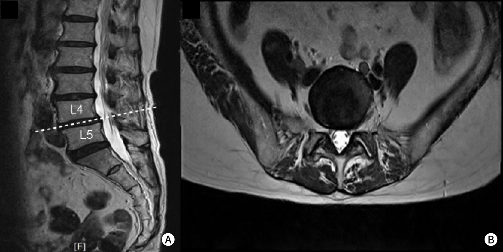
examinations were intact in both lower limbs. Lumbar MRI revealed multilevel
bulging discs and L4–5 degenerative spondylolisthesis, but no significant
neural foraminal stenosis or central canal stenosis (Fig. 1).
Fig. 1.
Lumbar MRI. (A) Sagittal view, (B) axial view.
Timeline
We initially diagnosed the patient with radiculopathy caused by degenerative
spondylolisthesis at L4–5, and therefore, a right L5–S1
transforaminal epidural steroid injection was performed. At a 1-week follow-up,
the patient’s pain score decreased from NRS 9 to 7 on the right lateral
thigh, but tingling and aching pain in the right buttock and posterior thigh
persisted. As the patient's pain was not effectively relieved, we
performed a piriformis muscle injection (0.1875% ropivacaine [5 mL],
dexamethasone [5 mg]) under ultrasound guidance to rule out piriformis syndrome.
After the procedure, she experienced gradual pain relief, with an NRS score of
5. We repeated the piriformis muscle injection 2 weeks later, but radiating pain
in the buttock and posterior thigh remained.
Diagnostic assessment
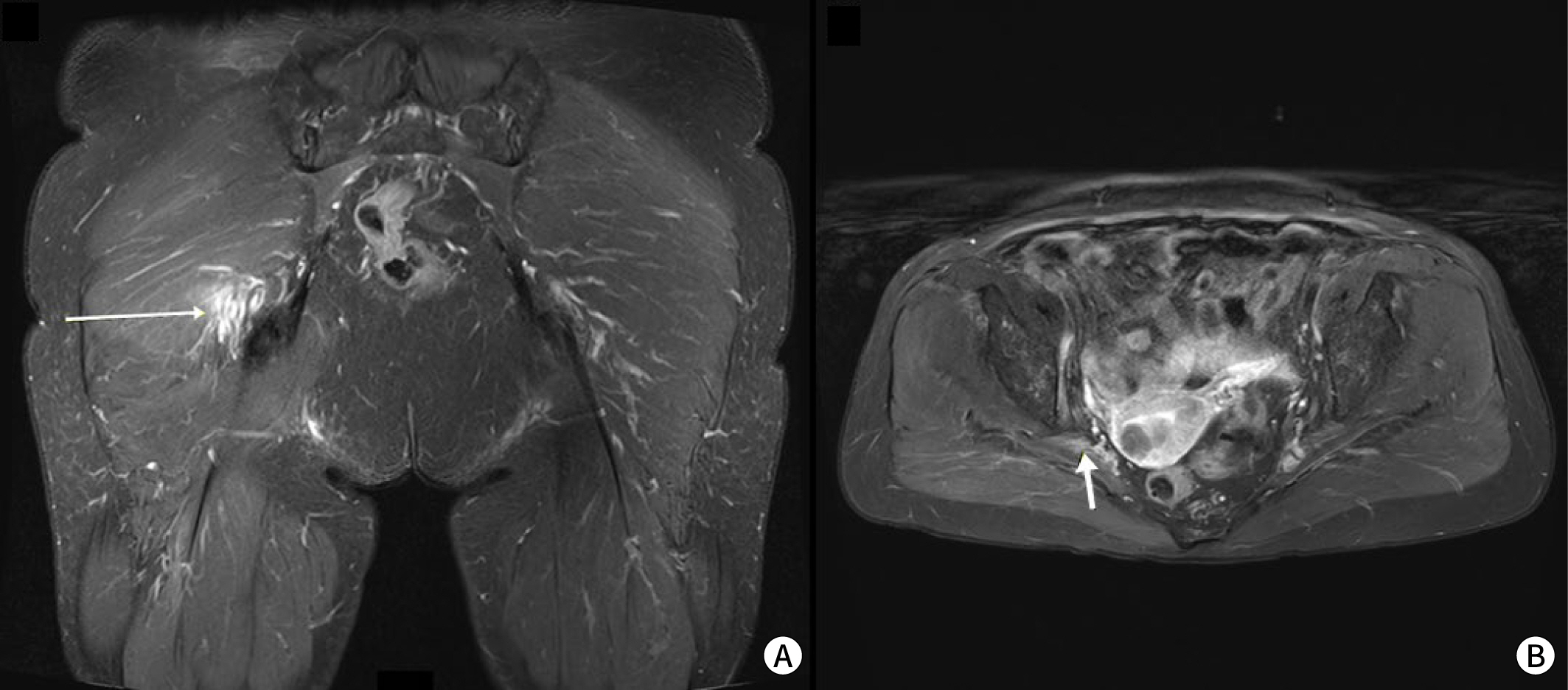
For further evaluation, we decided to perform hip MRI and a needle EMG study. EMG
did not reveal specific abnormal findings, but hip MRI showed thickening and T2
hyperintensity of the right sciatic nerve with perineural fat infiltrations
extending to the sub-gluteal region. Additionally, this area was adjacent to the
pedunculated portions of a uterine myoma (Fig.
2).
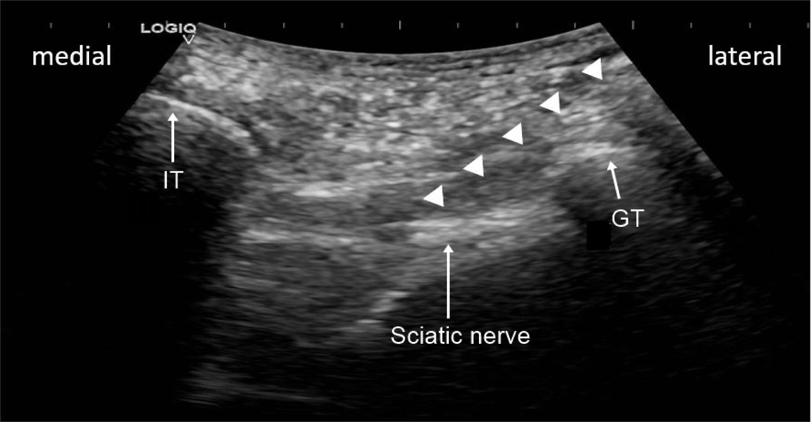
Based on the MRI findings, we planned to perform a sciatic nerve block under
ultrasound guidance. The patient was in a prone position. A curvilinear
transducer was then placed in the transverse plane at the lateral buttocks, and
the procedure was performed slightly below the gluteal region. The transducer
was placed between the greater trochanter and ischial tuberosity, and the
sub-gluteal space was seen as a hypoechoic space between the hyperechoic borders
of the gluteus maximus and quadratus femoris muscles, extending from the greater
trochanter to the ischial tuberosity. The sciatic nerve was visualized as an
oval hyperechoic structure between the greater trochanter and ischial
tuberosity. Under ultrasound guidance, we performed a sub-gluteal sciatic nerve
block with a mixture of 0.1875% ropivacaine (5 mL) and dexamethasone (5 mg;
Fig. 3).
The patient's pain was substantially relieved, reaching an NRS score of 2.
She expressed dramatic pain relief after the procedure and was totally
satisfied. No more aching symptoms remained when lying down or walking.
Following the procedure, she was prescribed a nonsteroidal anti-inflammatory
drug and antiepileptic drug for 4 weeks. At a follow-up visit 3 months later,
continued symptom improvement was noted. She was advised to return if her
symptoms worsened, but she did not attend any subsequent visits.
Discussion
To the best of the authors’ knowledge, this is the first report of successful
treatment using ultrasound-guided sciatic nerve block in a patient with sciatic
neuropathy objectively confirmed to have been caused by a uterine myoma based on hip
MRI.
Several cases have been reported in which a uterine myoma was identified as the cause
of sciatica [1,4–6]. In those cases,
despite variations in patient age, the size, number, and location of the myomas, and
proximity to menopause, the common treatment for sciatica was total hysterectomy.
Additionally, sciatic neuropathy was not objectively confirmed by MRI in those
cases. However, in our case, hysterectomy was not indicated, and we pursued an
alternative method to relieve her pain—specifically, based on the findings
from hip MRI, we performed an ultrasound-guided sciatic nerve block in the targeted
region.
Sciatic nerve block is categorized into parasacral, subgluteal, anterior, and
popliteal approaches based on the injection region. The target for the parasacral
approach is the sciatic nerve at a point distal to the lateral edge of the sacrum
and caudal to the sacroiliac joint [7].
However, this approach is essentially a sacral plexus block that targets branches of
the entire sacral plexus before the true sciatic nerve is formed at the inferior
edge of the piriformis muscle. It is performed at the level of the greater sciatic
foramen [7]. The sub-gluteal approach targets
the sciatic nerve as it traverses the sub-gluteal space, located between the greater
trochanter and the ischial tuberosity. This space is found between the posterior
aspect of the quadratus femoris and the anterior aspect of the gluteus maximus
[8,9]. In our patient, we performed a sub-gluteal sciatic nerve block at the
confirmed site under ultrasound guidance. The anterior approach aims at the sciatic
nerve in the proximal thigh as it descends medially to the femur, situated between
the adductor magnus anteriorly and the biceps femoris and semitendinosus
posteriorly. The popliteal approach targets the sciatic nerve as it divides into the
common peroneal nerve and the tibial nerve in the popliteal fossa region, typically
5–12 cm from the popliteal crease [9,10].
The sciatic nerve traverses a short intrapelvic course from the pelvis, passing
through the greater sciatic foramen [11]. The
sacral plexus is located on the posterior pelvic wall, anterior to the piriformis
muscle, and posterior to the sigmoid colon, ureter, and internal iliac vessels. Due
to its close proximity to the piriformis, the sciatic nerve is susceptible to
irritation and entrapment [12]. According to
a previous report, in cases of sciatica associated with obstetrical gynecological
disorders, endometriosis is the most common cause, followed by factors related to
pregnancy and labor, fibroids, sacral osteophytes, endosalpingiosis, needle
interventions, pelvic metastasis, piriformis-related sciatica, and singular cases
involving adenomyosis, intrauterine devices, hematocolpos, tubo-ovarian abscesses,
and retroverted uterus [13].
The treatment approach for uterine myomas is influenced by various considerations.
Relatively novel, less-invasive approaches are options, alongside pharmacological
therapy, conventional surgical methods, and expectant management. Surgical
procedures are considered if the patient exhibits abnormal uterine bleeding that
fails to respond to conservative management, or if there is strong suspicion of
pelvic malignancy, myoma growth after menopause, distortion of the endometrial
cavity in infertile women, pain or pressure symptoms that diminish quality of life,
or anemia resulting from chronic uterine blood loss [14]. In this case, our patient did not experience abnormal uterine
bleeding, but did report radiating lower extremity pain. She was not a candidate for
surgery; therefore, we planned an ultrasound-guided sciatic nerve block.
It is challenging to accurately explain why pain relief was achieved after just one
sciatic nerve block without treating the causative disease. Even a single nerve
block using a local anesthetic can provide lasting pain relief, which is attributed
to neuroplasticity [15]. This likely applies
to our case as well. Additionally, this patient underwent piriformis muscle
injections twice before the sub-gluteal sciatic nerve block. During piriformis
muscle injections, the medication can spread to the sciatic nerve, leading to a
parasacral sciatic nerve block. This may explain why the patient's symptoms
were somewhat alleviated after the piriformis muscle injections. In other words,
although the patient only underwent one sub-gluteal sciatic nerve block, it can be
considered that she experienced repeated sciatic nerve blocks as a result of the
spread of medication during the previous injections.
In conclusion, when female patients experience radiating pain that does not respond
as expected to lumbar treatment, it is crucial to consider the possibility of
gynecologic problems in the differential diagnosis of sciatica. Hip MRI can be
helpful for diagnosis, and if surgical treatment is not indicated, ultrasound-guided
sciatic nerve block can be an appropriate and effective treatment option.
Authors' contributions
Project administration: Kim W
Conceptualization: Kim W
Methodology & data curation: Kang BK, Beak MH
Funding acquisition: not applicable
Writing – original draft: Kang BK, Beak MH, Kim W
Writing – review & editing: Kang BK, Beak MH, Kim W
Conflict of interest
No potential conflict of interest relevant to this article was reported.
Funding
Not applicable.
Data availability
Not applicable.
Acknowledgments
Not applicable.
Supplementary materials
Not applicable.
References
1. Bodack MP, Cole JC, Nagler W. Sciatic neuropathy secondary to a uterine fibroid: a case
report. Am J Phys Med Rehabil 1999;78(2):157-159.
8. Karmakar MK, Kwok WH, Ho AM, Tsang K, Chui PT, Gin T. Ultrasound-guided sciatic nerve block: description of a new
approach at the subgluteal space. Br J Anaesth 2007;98(3):390-395.
12. Beaton LE, Anson BJ. The sciatic nerve and the piriformis muscle: their interrelation
a possible cause of coccygodynia. J Bone Joint Surg 1938;20(3):686-688.
13. Khodairy AW, Bovay P, Gobelet C. Sciatica in the female patient: anatomical considerations,
aetiology and review of the literature. Eur Spine J 2007;16(6):721-731.
AVANÇOS E PERSPECTIVAS NA ANESTESIA NEUROAXIAL: SEGURANÇA, EFICÁCIA E APLICAÇÕES CLÍNICAS MODERNAS Gabriel Fontes , Julia de Souza e Silva, Gabriel Vitor Ferreira, Sofia Boechat Melado, Guilherme Soares Carvalho, Julia Kalene Saraiva Torres, Leonardo Lanes Leite Silvestre, Luís Pedro Cerqueira Morejón, Laryssa Inácio Carvalho, Matheus dos Santos Nunes, Revista Contemporânea.2025; 5(1): e7282. CrossRef