1Division of Gastroenterology, Pusan National University Hospital, Busan, Korea
2Department of Internal Medicine, Pusan National University School of Medicine, Busan, Korea
3Biomedical Research Institute, Pusan National University Hospital, Busan, Korea
4Department of Pathology, Pusan National University Hospital, Busan, Korea
*Corresponding author: Gwang Ha Kim,
Department of Internal Medicine, Pusan National University School of Medicine,
and Biomedical Research Institute, Pusan National University Hospital, 179
Gudeok-ro, Seo-gu, Busan 49241, Korea E-mail:
doc0224@pusan.ac.kr
• Received: March 4, 2024 • Revised: April 19, 2024 • Accepted: April 19, 2024
This is an Open-Access article distributed under the terms of the
Creative Commons Attribution Non-Commercial License (http://creativecommons.org/licenses/by-nc/4.0) which permits
unrestricted non-commercial use, distribution, and reproduction in any
medium, provided the original work is properly cited.
We report a rare case of gastric adenocarcinoma with enteroblastic
differentiation (GAED) that was treated with endoscopic submucosal dissection
followed by additional distal gastrectomy with lymph node dissection. A
67-year-old man underwent endoscopic submucosal dissection for a gastric lesion,
which was diagnosed as GAED with submucosal and lymphatic invasion.
Histologically, GAED is characterized by a tubulopapillary growth pattern and
clear cells that resemble those of the primitive fetal gut.
Immunohistochemically, GAED variably expresses oncofetal proteins such as
glypican-3, alpha-fetoprotein, and spalt-like transcription factor 4. Despite
negative margins, additional gastrectomy with lymph node dissection was
performed due to submucosal and lymphatic invasion. No residual tumor or
metastasis was detected, and the patient remained disease-free for 2 years
before dying from causes unrelated to GAED. Given its aggressive nature,
frequent lymphovascular invasion, and high metastatic potential, clinicians
should recognize the histopathological diagnosis of this rare tumor and its
propensity for aggressiveness.
Gastric adenocarcinoma with enteroblastic differentiation (GAED), also known as clear
cell gastric carcinoma, is a rare and poorly documented malignancy, representing
less than 1% of all gastric cancers [1,2]. While GAED represents a subtype of
alpha-fetoprotein (AFP)-producing adenocarcinomas [1], the relationship between GAED and AFP production is not well
understood [2]. Histologically, the tumor is
characterized by an intestine-like structure composed of cuboidal or columnar
neoplastic cells with clear cytoplasm. These cells test positive for oncofetal
proteins, such as glypican-3, spalt-like transcription factor 4 (SALL4), and AFP
[3]. GAED tends to be more aggressive than
conventional adenocarcinoma, with a higher propensity for lymphovascular invasion
and a greater likelihood of metastasis to the liver and lymph nodes (LNs) [4]. In this report, we present a rare case of
GAED that was managed with endoscopic submucosal dissection and additional distal
gastrectomy with LN dissection.
Case presentation
Ethics statement
This case report was granted an exemption from consent and review by the Pusan
National University Hospital Research Ethics Review Committee (IRB No.
2402-023-136).
Patient information
A 67-year-old man visited our hospital seeking treatment for high-grade dysplasia
in the stomach, which was identified during esophagogastroduodenoscopy at a
health checkup. The patient was asymptomatic. His medical history included
alcoholic hepatitis and chronic hepatitis B, along with heavy alcohol
consumption and a 40-pack-year smoking history.
Clinical findings
The results of the physical examination were unremarkable.
Diagnostic assessment
Laboratory analysis indicated a slight elevation of liver function test results,
suggestive of alcoholic hepatitis. Tumor markers, including serum AFP,
carcinoembryonic antigen, and carbohydrate antigen 19–9, were within
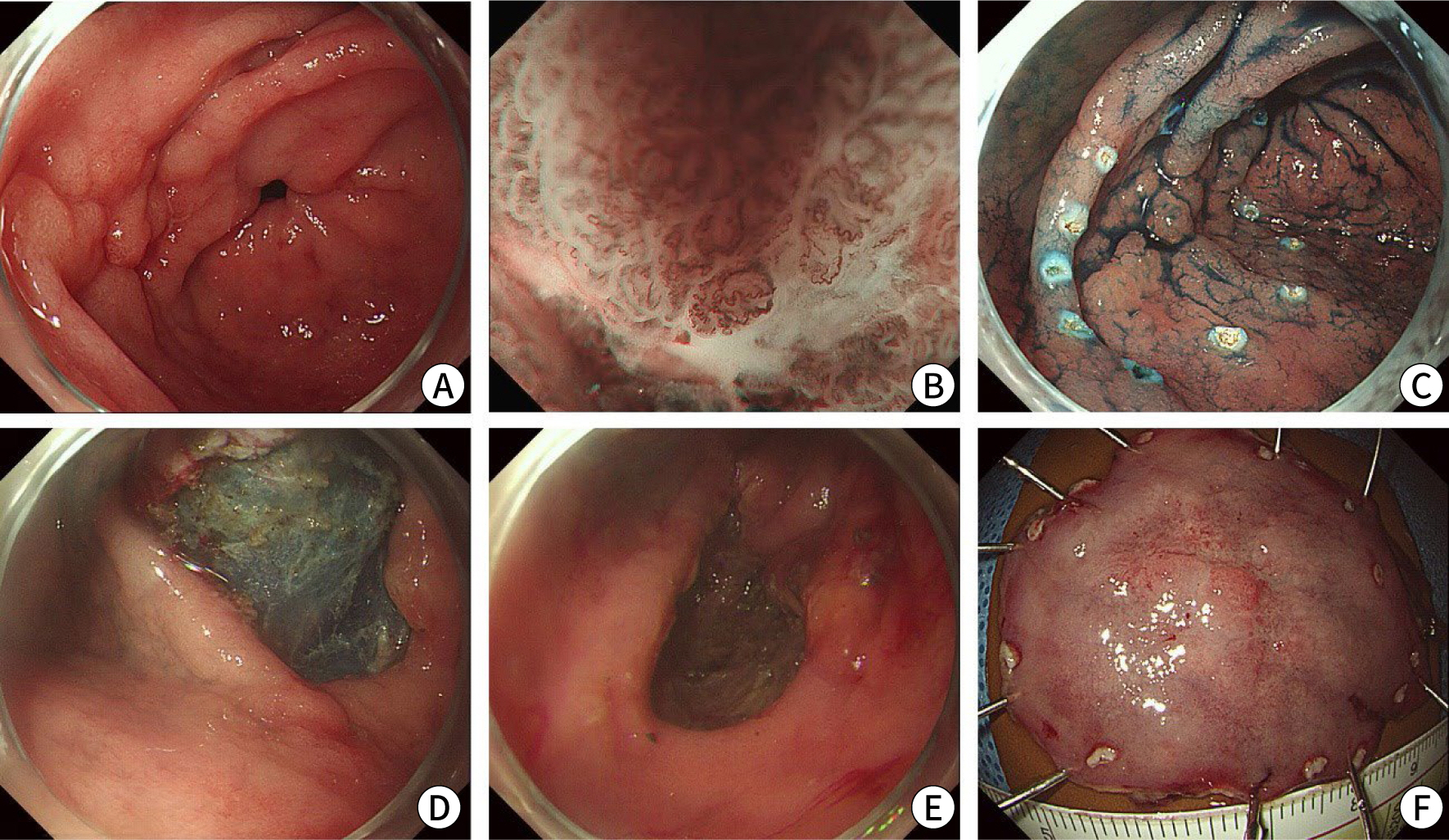
normal limits. Esophagogastroduodenoscopy revealed a 2-cm slightly depressed
lesion with nodular mucosal changes on the anterior wall of the gastric
prepylorus (Fig. 1A). Magnifying endoscopy
with narrow-band imaging revealed a clear demarcation line and irregular
patterns in microsurface (MS) and microvascular (MV) structures, including
irregular oval/tubular MS and irregular loop MV patterns (Fig. 1B). Endoscopic ultrasonography indicated that the
lesion was confined to the mucosal layer. Abdominal and chest computed
tomography revealed no evidence of LN involvement or distant metastases.
Fig. 1.
Endoscopic submucosal dissection for early gastric cancer. (A)
Conventional endoscopy and indigo carmine chromoendoscopy reveal a 2-cm
slightly depressed lesion with nodular mucosal changes on the anterior
wall of the gastric prepylorus. (B) Magnifying endoscopy with
narrow-band imaging shows irregular microsurface and microvascular
patterns. (C) Marking dots are placed around the lesion. (D) A
circumferential incision and submucosal dissection are performed using
an insulated-tip knife. (E) The lesion is completely excised. (F) The
resected specimen is shown.
Therapeutic intervention and final diagnosis
Endoscopic submucosal dissection was performed to achieve complete resection of
the lesion (Fig. 1C–E). Assessment of the gross appearance of the
resected specimen revealed a 19-mm, IIc lesion with an irregular mucosal surface
(Fig. 1F). Based on microscopic
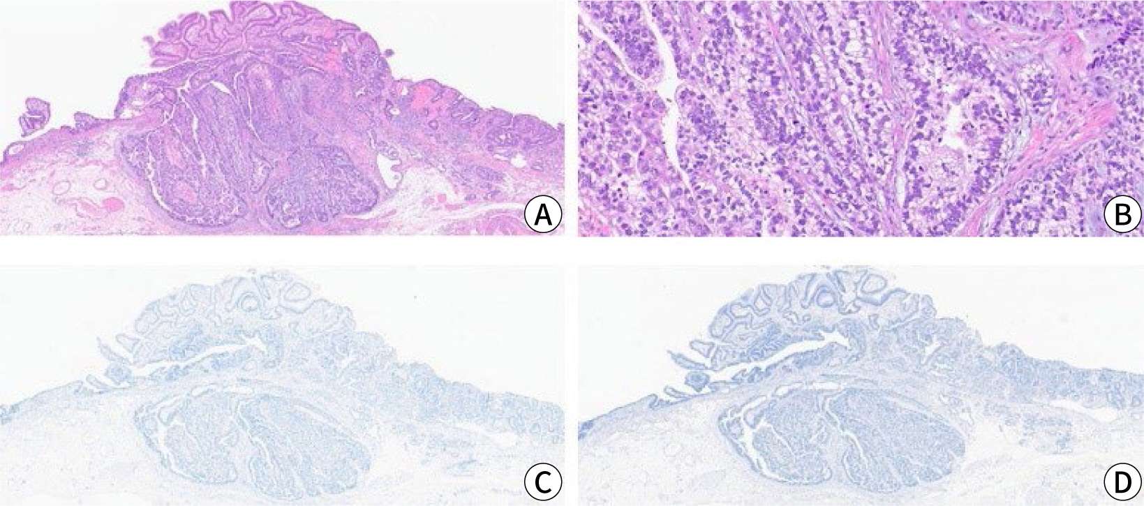
examination, the tumor exhibited a tubulopapillary growth pattern along with
submucosal invasion (Fig. 2A). The tumor
was overlaid by conventional adenocarcinoma and was partially composed of
cuboidal or columnar cells with clear cytoplasm, a feature reminiscent of the
primitive fetal gut and indicative of enteroblastic differentiation (Fig. 2B). On immunohistochemical staining,
the tumor cells tested negative for glypican-3 and AFP, which are recognized as
oncofetal proteins (Fig. 2C, D). Although the horizontal and deep
resection margins were tumor-free, the tumor had penetrated the deep submucosa
(750 µm from the muscularis mucosa) and exhibited lymphatic invasion.
Consequently, additional distal gastrectomy with LN dissection was performed.
Examination of the surgical specimens revealed no residual tumor or LN
metastasis.
Fig. 2.
Histopathological findings. (A) The tumor exhibits a tubulopapillary
growth pattern and submucosal invasion (hematoxylin and eosin
[H&E] stain, ×40). (B) Tumor cells display clear cytoplasm
and a tubular pattern, indicative of enteroblastic adenocarcinoma
(H&E stain, ×200). (C,D) Tumor cells test negative for
glypican-3 (C) and alpha-fetoprotein (D) (immunohistochemical stain,
×40).
Follow-up and outcomes
During the 2-year follow-up period, we observed no evidence of local or distant
recurrence. However, 3 years after the intervention for GAED, the patient died
of necrotizing pneumonia and uncontrolled alcoholic hepatitis.
Discussion
GAED, also known as clear cell gastric carcinoma, is a rare entity in the stomach.
Clear cell carcinomas are more commonly found in the lower urinary tract and the
female reproductive system, specifically the endometrium and ovary. Due to the
infrequency of GAED, its clinicopathologic and immunohistochemical features are not
yet fully understood [2,5,6]. Histologically,
enteroblastic adenocarcinoma often coexists with conventional well-differentiated or
moderately-differentiated tubular adenocarcinoma, found in the upper portion of the
tumor [2]. GAED is characterized by a
tubulopapillary growth pattern, with predominantly clear cells and luminal
eosinophilic secretions [7]. In the present
case, magnifying endoscopy with narrow-band imaging revealed irregular oval/tubular
MS and irregular loop MV patterns. These findings were in line with the
histopathological characteristics of GAED, as described in previous reports [8,9].
On immunohistochemical staining, most GAEDs variably express three enteroblastic
lineage markers, also known as oncofetal proteins. These markers include glypican-3
(associated with hepatoid gastric carcinoma), AFP (a marker for hepatocellular
carcinoma and yolk sac tumor), and SALL4 (a marker for AFP-producing gastric
carcinoma) [10−12]. For the diagnosis of GAED, glypican-3 is the most
sensitive marker, followed by SALL4 and AFP [2]. In the present case, staining was negative for glypican-3 and AFP, and
SALL4 staining could not be performed due to a lack of necessary testing equipment.
When immunohistochemical staining reveals AFP production in gastric carcinoma cells,
the lesion is classified as AFP-producing gastric carcinoma. This type of carcinoma
is associated with a poor prognosis due to a high incidence of lymphovascular
invasion and liver metastasis [13].
Clear cells are characterized by an abundant cytoplasm filled with substances such as
glycogen, lipid, water, or mucin [6]. Gastric
carcinomas with clear cell changes (GCCs) typically display a cytoplasmic
accumulation of glycogen and mucin. Our previous research indicated that GCCs
secondary to glycogen deposition are associated with the expression of AFP,
glypican-3, and CD10. In contrast, GCCs with mucin deposition are linked to the
expression of MUC5AC and MUC6 [14]. GAED and
hepatoid adenocarcinoma are representative histologic subtypes of GCC. Hepatoid
adenocarcinoma with clear cells is distinguished from GAED by its poor prognosis,
diffuse and strong expression of oncofetal proteins, and intestinal mucin phenotype
[7]. In contrast, GAED exhibits focally
heterogenous expression of oncofetal proteins and frequently expresses CD10, CDX-2,
and MUC6, but not MUC2 and MUC5AC. These features suggest a gastric
antral/intestinal mucin phenotype with focal enteroblastic differentiation [7].
Similar to AFP-producing adenocarcinoma, the presence of clear cell changes in
gastric cancer is associated with a poor prognosis compared to conventional gastric
adenocarcinoma [14]. Research indicates that
most patients with GAED (90%) exhibit lymphatic and/or vascular invasion [2]. LN metastasis is observed in 40% of
early-stage cases and 84% of advanced cases, which exceeds the rates observed in
conventional gastric adenocarcinoma (20%–45%).
In conclusion, GAED is a rare malignancy characterized by distinct histopathological
features. It is more aggressive than conventional adenocarcinoma, with frequent
lymphovascular invasion and metastasis to the liver and LNs; consequently, it has a
poor prognosis. Clinicians should therefore recognize the histopathological
diagnosis of this rare tumor and remain cognizant of its aggressive behavior.
Authors' contributions
Project administration: Kim GH
Conceptualization: Kim GH
Methodology & data curation: Joo DC, Lee MW, Lee BE, Kim KB
Funding acquisition: not applicable
Writing – original draft: Lee HR, Kim GH
Writing – review & editing: Lee HR, Kim GH, Joo DC, Lee MW, Lee BE,
Kim KB
Conflict of interest
No potential conflict of interest relevant to this article was reported.
Funding
Not applicable.
Data availability
Not applicable.
Acknowledgments
Not applicable.
Supplementary materials
Not applicable.
References
1. Kumar S, Jabbar K. Gastric adenocarcinoma with enteroblastic differentiation: a rare
find. Am J Clin Pathol 2020;154(Suppl 1):S65
2. Murakami T, Yao T, Mitomi H, Morimoto T, Ueyama H, Matsumoto K, et al. Clinicopathologic and immunohistochemical characteristics of
gastric adenocarcinoma with enteroblastic differentiation: a study of 29
cases. Gastric Cancer 2016;19(2):498-507.
3. Dias E, Santos-Antunes J, Nunes AC, Rodrigues JA, Pinheiro J, Macedo G. Gastric adenocarcinoma with enteroblastic differentiation: an
unexpected cause of upper gastrointestinal bleeding. Acta Gastroenterol Belg 2021;84(4):678-679.
4. Abada E, Anaya IC, Abada O, Lebbos A, Beydoun R. Colorectal adenocarcinoma with enteroblastic differentiation:
diagnostic challenges of a rare case encountered in clinical
practice. J Pathol Transl Med 2022;56(2):97-102.
5. Afshar Ghotli Z, Serra S, Chetty R. Clear cell (glycogen rich) gastric adenocarcinoma: a distinct
tubulo‐papillary variant with a predilection for the
cardia/gastro‐oesophageal region. Pathology 2007;39(5):466-469.
7. Kwon MJ, Byeon S, Kang SY, Kim KM. Gastric adenocarcinoma with enteroblastic differentiation should
be differentiated from hepatoid adenocarcinoma: a study with emphasis on
clear cells and clinicopathologic spectrum. Pathol Res Pract 2019;215(9):152525
8. Ishikawa A, Nakamura K. Gastric adenocarcinoma with enteroblastic differentiation
resected through endoscopic submucosal dissection: a case
report. Case Rep Gastroenterol 2024;18(1):68-73.
9. Kato T, Hikichi T, Nakamura J, Takasumi M, Hashimoto M, Kobashi R, et al. Two cases of gastric adenocarcinoma with enteroblastic
differentiation resected by endoscopic submucosal dissection. Clin J Gastroenterol 2021;14(3):736-744.
10. Kinjo T, Taniguchi H, Kushima R, Sekine S, Oda I, Saka M, et al. Histologic and immunohistochemical analyses of
α-fetoprotein: producing cancer of the stomach. Am J Surg Pathol 2012;36(1):56-65.
11. Ushiku T, Shinozaki A, Shibahara J, Iwasaki Y, Tateishi Y, Funata N, et al. SALL4 represents fetal gut differentiation of gastric cancer, and
is diagnostically useful in distinguishing hepatoid gastric carcinoma from
hepatocellular carcinoma. Am J Surg Pathol 2010;34(4):533-540.
12. Yamauchi N, Watanabe A, Hishinuma M, Ohashi KI, Midorikawa Y, Morishita Y, et al. The glypican 3 oncofetal protein is a promising diagnostic marker
for hepatocellular carcinoma. Mod Pathol 2005;18(12):1591-1598.
14. Kim JY, Park DY, Kim GH, Jeon TY, Lauwers GY. Does clear cell carcinoma of stomach exist? Clinicopathological
and prognostic significance of clear cell changes in gastric
adenocarcinomas. Histopathology 2014;65(1):90-99.
Gastric adenocarcinoma with enteroblastic differentiation in a
67-year-old man in Korea: a case report
Fig. 1.
Endoscopic submucosal dissection for early gastric cancer. (A)
Conventional endoscopy and indigo carmine chromoendoscopy reveal a 2-cm
slightly depressed lesion with nodular mucosal changes on the anterior
wall of the gastric prepylorus. (B) Magnifying endoscopy with
narrow-band imaging shows irregular microsurface and microvascular
patterns. (C) Marking dots are placed around the lesion. (D) A
circumferential incision and submucosal dissection are performed using
an insulated-tip knife. (E) The lesion is completely excised. (F) The
resected specimen is shown.
Fig. 2.
Histopathological findings. (A) The tumor exhibits a tubulopapillary
growth pattern and submucosal invasion (hematoxylin and eosin
[H&E] stain, ×40). (B) Tumor cells display clear cytoplasm
and a tubular pattern, indicative of enteroblastic adenocarcinoma
(H&E stain, ×200). (C,D) Tumor cells test negative for
glypican-3 (C) and alpha-fetoprotein (D) (immunohistochemical stain,
×40).
Fig. 1.
Fig. 2.
Gastric adenocarcinoma with enteroblastic differentiation in a
67-year-old man in Korea: a case report

 , Gwang Ha Kim1,2,3,*