1Department of Anesthesiology and Pain Medicine, Ewha Womans University College of Medicine, Seoul, Korea
2Department of Anesthesiology and Pain Medicine, Ewha Womans University Mokdong Hospital, Ewha Womans University College of Medicine, Seoul, Korea
*Corresponding author: Sooyoung Cho,
Department of Anesthesiology and Pain Medicine, Ewha Womans University Mokdong
Hospital, Ewha Womans University College of Medicine, 1071 Anyangcheon-ro,
Yangcheon-gu, Seoul 07985, Korea E-mail:
sooyoung.cho@ewha.ac.kr
• Received: December 1, 2023 • Revised: January 20, 2024 • Accepted: April 17, 2024
This is an Open-Access article distributed under the terms of the
Creative Commons Attribution Non-Commercial License (http://creativecommons.org/licenses/by-nc/4.0) which permits
unrestricted non-commercial use, distribution, and reproduction in any
medium, provided the original work is properly cited.
OxyMask, a novel product, has recently been used to administer oxygen
postoperatively to patients who have undergone general anesthesia. This
study aimed to evaluate the incidence of hypoxia in patients under general
anesthesia upon arrival to the post-anesthesia care unit (PACU) using
arterial blood gas analysis, and to compare the effectiveness of OxyMask
with a non-rebreathing oxygen mask for oxygen administration.
Methods:
We retrospectively investigated anesthesia-related data from the electronic
medical records of 460 patients treated from April to November 2021. We
analyzed patients aged 20 years or older who had undergone general
anesthesia and whose perioperative arterial blood gas analysis results were
available upon arrival to the PACU. These patients were grouped into the
non-rebreathing oxygen mask (n=223) and OxyMask (n=237) groups, and
statistical analysis was performed utilizing their anesthesia records.
Results:
No patients exhibited hypoxia upon arrival to the recovery room. The oxygen
concentration increased after oxygen administration; its concentration
during the recovery room period (Δ2 PaO2) was
10.7±42.3 and 13.9±38.5 mmHg in the non-rebreathing oxygen
mask and OxyMask groups, respectively. This difference was not statistically
significant. Moreover, the arterial oxygen saturation between the end of
surgery and upon arrival to the PACU (Δ1 SaO2) and the
arterial oxygen saturation 20 minutes after oxygen administration at the
PACU (Δ2 SaO2) did not significantly differ between the
groups.
Conclusion:
OxyMask was not superior to a non-rebreathing oxygen mask in terms of the
effectiveness of oxygen supply.
The immediate postoperative period is a risky time when hypoxia is highly likely
to occur. Should respiratory complications arise during post-anesthesia
management, they can lead to serious consequences. Thus, comprehensive
management is essential to ensure thorough monitoring of the patient.
Postoperative hypoxia, which develops immediately after surgery, is
significantly associated with factors such as anesthesia duration, surgical
incision site, age, obesity, and pain [1–5].
All general patients are transferred from the operating room (OR) to the
post-anesthesia care unit (PACU) after surgery, breathing room air; at this
point, there is a risk of developing hypoxia. Upon arrival in the PACU, patients
receive oxygen through various devices such as masks and cannulas. Previously,
our institution administered oxygen using a non-rebreathing oxygen mask
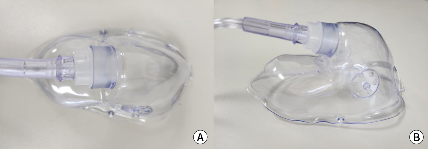
(Teleflex, Morrisville, NC, USA; Fig. 1);
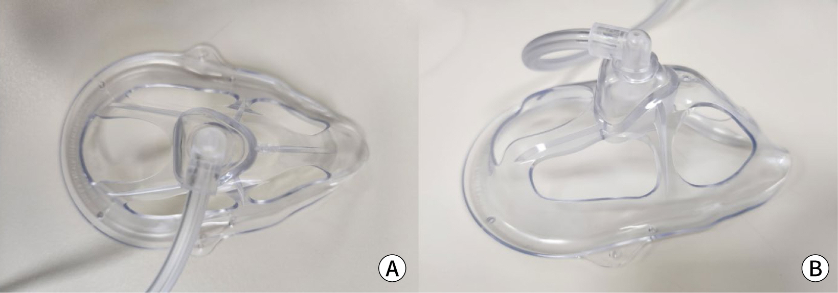
however, we have recently begun using the OxyMask (Southmedic, Barrie, ON,
Canada), a novel product (Fig. 2).
Fig. 1.
Hudson RCI non-rebreathing oxygen mask. (A) Top view, (B) side
view.
Fig. 2.
OxyMask. (A) Top view, (B) side view.
A non-rebreathing oxygen mask is a closed-type mask, which leads to air
re-circulation at the lower part of the mask, specifically around the chin area.
This can result in the accumulation of carbon dioxide (CO2) in the
expiratory gas [6]. The flow of oxygen in
this mask is almost parallel to the face and directed toward the nose, which may
not effectively support oxygen inhalation through the mouth. In contrast, the
OxyMask is an open-type mask designed to prevent the accumulation of
CO2 inside the mask by allowing oxygen to flow from the center of
the mask towards both the nasal and oral cavities [6]. This design could facilitate oxygen inhalation and
improve CO2 removal from the mask. However, there is a concern that
the open structure of the mask might lead to oxygen dispersion.
Objectives
We assessed the incidence and severity of hypoxia during patient transfer from
the OR to the PACU in individuals who had undergone general anesthesia and were
extubated. Additionally, this study compared the effectiveness of a
non-rebreathing oxygen mask and OxyMask in the PACU by utilizing arterial blood
gas analysis (ABGA).
Methods
Ethics statement
The study was conducted in accordance with the Declaration of Helsinki and
approved by the Institutional Review Board (IRB) of Ewha Womans University
Mokdong Hospital (IRB number: 2021-12-048-001). The requirement to obtain
written informed consent was waived by the IRB since this study was performed
retrospectively.
We reviewed and analyzed patients’ data from their electronic medical
records from April to November 2021 at Ewha Womans Medical Center. A
non-rebreathing oxygen mask was used from May 1, 2021 to July 31, 2021. The
OxyMask was used from August 1, 2021 to November 30, 2021.
Participants
We investigated 460 patients aged 20 years or older who had undergone general
anesthesia in non-cardiac surgery, were extubated at the end of anesthesia, and
had perioperative ABGA results available upon arrival to the PACU and before
leaving the PACU. Patients with severe cardiopulmonary disease who experienced
hypoxia and dyspnea and were therefore administered supplemental oxygen from the
OR were excluded. The general protocol for anesthesia at our institution was as
follows: Perioperatively, the management of anesthesia was at the discretion of
the attending anesthesiologist. All patients were extubated in the OR and then
transferred to the PACU after confirming spontaneous breathing with an oxygen
supply via a bag-mask at 6 L/min for at least 5 minutes. During the transfer to
the PACU, all patients breathed room air (FiO2 0.21) spontaneously.
Upon arrival to the PACU, all patients received supplemental oxygen at 6 L/min
(FiO2 0.44) using a non-rebreathing oxygen mask or an OxyMask,
and an ABGA was performed (the first ABGA in the PACU). After at least 20
minutes of oxygen administration in the PACU, a second ABGA was performed for
all patients. The attending anesthesiologist determined whether to discharge
patients from the PACU based on the discharge criteria, following the Aldrete
score system, which evaluates activity, respiration, circulation, consciousness,
and skin color (Supplement 1) after discontinuation of the oxygen supply.
Variables (study outcomes)
The primary outcome was a comparison of the effectiveness of oxygen delivery
between the OxyMask and the non-rebreathing oxygen mask during the stay in the
PACU, as determined by ABGA results. Postoperative hypoxia was defined as an
oxyhemoglobin saturation (SpO2) of 90% or less for at least 2
minutes, or an SpO2 of 85% or less at any time point. The secondary
outcome focused on the incidence of postoperative hypoxia upon arrival to the
PACU, which was also assessed using ABGA results.
Data sources and measurement
The collected data included the patients’ demographic and clinical
characteristics, as follows: sex, age, American Society of Anesthesiologists
physical status, height, weight, body mass index, the presence of pulmonary
disease, operation time, and anesthesia time. Intraoperative and postoperative
vital signs and ABGA results were collected, and the following vital signs were
recorded at the time of ABGA sampling: systolic blood pressure, diastolic blood
pressure, heart rate, respiratory rate, SpO2, respiratory pattern,
level of consciousness, and numerical pain scores were recorded every 5 minutes
in all patients. From the ABGA results, pH, arterial oxygen partial pressure
(PaO2), arterial carbon dioxide partial pressure, and arterial
oxygen saturation (SaO2) were also recorded. Additionally, we
calculated the difference in PaO2 and SaO2 between the
last ABGA in the OR and the first ABGA in the PACU (Δ1
PaO2 and Δ1 SaO2, respectively).
Moreover, we calculated the difference in PaO2 and SaO2
between the first and second PACU measurements (Δ2
PaO2 and Δ2 SaO2, respectively).
Bias
There was no selection bias reportable in this study.
Study size
Sample size estimation was not performed because this study included all target
patients, with the exclusion of those who received supplemental oxygen in the
OR. Additionally, the authors did not allocate participants to specific
groups.
Statistical methods
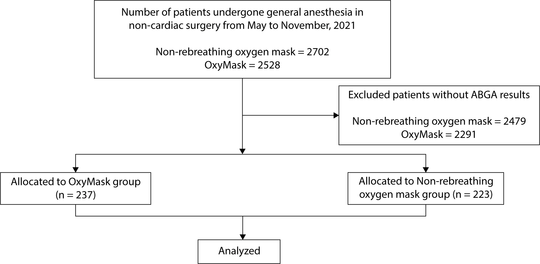
Statistical analyses were performed using the anesthesia records of 460 patients,
divided into two groups: the OxyMask group (n=237) and the non-rebreathing
oxygen mask group (n=223; Fig. 3).
Continuous variables were analyzed using either the Student t-test or the
Mann–Whitney U test following a normality assessment with the
Shapiro–Wilk test. Results were presented as means±SDs or as
medians (interquartile ranges). Categorical variables were analyzed using the
chi-square test or Fisher exact test, which was applied when more than 20% of
the expected frequencies were fewer than 5. These results were presented as
percentages (%). All statistical analyses were carried out using SPSS version 25
(IBM, Armonk, NY, USA). A P-value of less than 0.05 was considered statistically
significant.
Fig. 3.
Flow diagram of the participants.
Results
Participants’ demographic and clinical characteristics in the
non-rebreathing oxygen mask and OxyMask groups
We analyzed 460 patients, who were divided into the OxyMask group (n=237) and
non-rebreathing oxygen mask group (n=223). The two groups did not differ
significantly in terms of demographic or clinical characteristics, except the
operation time (167.9±125.2 vs. 192.9±139.8 min, P=0.044; Table 1). Patients who had pulmonary
disease were comparable between the OxyMask and non-rebreathing oxygen mask
groups (Table 1).
Table 1.
Demographic characteristics of both groups
Demographic characteristic
Non-rebreathing oxygen mask
(n=223)
OxyMask (n=237)
P-value
Age (years)
60.6±17.3
62.3±15.4
0.275
Sex (M/F)
91/132
105/132
0.448
Height (cm)
160.6±9.9
162.2±9.1
0.064
Weight (kg)
60.6±11.9
63.4±11.0
0.010
BMI (kg/m2)
23.5±3.8
24.1±3.7
0.073
ASA physical classification
(1/2/3/4)
41/137/45/0
27/154/55/1
0.141
Pulmonary disease (yes/no)
28/195
39/198
0.236
Operation time (min)
167.9±125.2
192.9±139.8
0.044
Anesthesia time (min)
215.4±130.3
237.8±141.8
0.079
Values are presented as mean±SDs.
The Student t-test was performed for continuous variables and the
chi-square test for categorical variables.
P<0.05 is regarded as indicating statistical significance.
BMI, body mass index; ASA, American Society of Anesthesiologists.
Incidence of postoperative hypoxia upon arrival to the post-anesthesia care
unit
No hypoxia episodes occurred among the patients. One patient in the OxyMask group
had a minimum PaO2 value of 111.8 mmHg upon arrival to the PACU, with
spontaneous breathing of room air after surgery.
Arterial blood gas analysis during the operation and the post-anesthesia care
unit period
Upon arrival to the PACU, PaO2 was significantly lower in the OxyMask
group than in the non-rebreathing oxygen mask group (162.1±50.3 vs.
181.9±62.0 mmHg, respectively, P<0.001). Similarly, upon discharge
from the PACU, PaO2 was significantly lower in the OxyMask group than
in the non-rebreathing oxygen mask group. (176.0±49.2 vs.
192.6±64.5 mmHg, respectively, P=0.002).
Δ1 PaO2, the gradient of arterial oxygen partial
pressure between the end of surgery and upon arrival to the PACU, was
significantly higher in the OxyMask group than in the non-rebreathing oxygen
mask group (40.5±48.7 vs. 19.9±52.8 mmHg, respectively,
P<0.001; Table 2). However,
Δ2 PaO2, the gradient of arterial oxygen
partial pressure 20 minutes after the administration of oxygen in the PACU, was
not significantly different between the OxyMask and the non-rebreathing oxygen
mask groups (13.9±38.5 vs. 10.7±42.3 mmHg, respectively, P=0.393;
Table 2).
Table 2.
Arterial blood gas analysis during operation and the PACU
period
Variable
During the operation
(OR)
Arrival at the PACU
(PACU1)
Discharge from the PACU
(PACU2)
Non-rebreathing oxygen mask
(n=223)
OxyMask (n=237)
P-value
Non-rebreathing oxygen mask
(n=223)
OxyMask (n=237)
P-value
Non-rebreathing oxygen mask
(n=223)
OxyMask (n=237)
P-value
SBP
117.8±17.1
119.4±16.9
0.310
129.8±8.3
133.5±21.3
0.049
129.6±18.6
134.6±21.7
0.009
DBP
66.6±12.0
66.3±11.4
0.807
77.1±14.3
79.3±16.3
0.115
75.7±11.6
77.8±14.2
0.095
HR
79.7±14.1
80.3±14.7
0.662
85.4±13.9
85.0±15.1
0.768
79.7±13.4
80.9±14.3
0.362
PACU
13.2±4.4
12.9±4.3
0.471
17.7±4.1
17.6±6.8
0.769
16.6±6.2
16.4±3.7
0.686
SpO2
100.0±0.3
100.0±0.4
0.941
100.0±0.0
100.0±0.2
0.207
100.0±0.0
100.0±0.0
-
pH
7.40±0.05
7.41±0.05
0.211
7.38±0.05
7.38±0.05
0.719
7.39±0.06
7.40±0.05
0.699
pCO2
38.3±3.6
37.8±3.4
0.106
40.0±5.4
39.3±5.4
0.153
38.1±5.3
37.4±4.8
0.155
pO2
201.8±48.4
202.6±47.6
0.850
181.9±62.0
162.1±50.3
<0.001*
192.6±64.5
176.0±49.2
0.002*
Δ1PaO2
-
-
-
19.9±52.8
40.5±48.7
<0.001*
-
-
-
Δ2PaO2
-
-
-
-
-
-
10.7±42.3
13.9±38.5
0.393
SaO2
98.7±0.7
98.6±0.8
0.256
98.0±5.0
98.1±1.2
0.736
98.5±0.7
98.5±0.7
0.193
Δ1SaO2
-
-
-
0.13±0.70
0.14±0.77
0.912
-
-
-
Δ2SaO2
-
-
-
-
-
-
0.51±4.93
0.32±1.02
0.544
Values are presented as mean±SD.
The Student t-test was performed for continuous variables.
P<0.05 is regarded as indicating statistical significance, and
* significant results are shown.
OR, operating room; PACU, post-anesthesia care unit; SBP, systolic
blood pressure; DBP, diastolic blood pressure; HR, heart rate; PACU,
respiratory rate; SpO2, oxyhemoglobin saturation;
PaCO2, arterial carbon dioxide partial pressure;
PaO2, arterial oxygen partial pressure;
Δ1PaO2, PaO2 at
OR–PaO2 at PACU1;
Δ2PaO2, PaO2 at
PACU2–PaO2 at PACU1;
SaO2, arterial oxygen saturation;
Δ1SaO2, SaO2 at
OR–SaO2 at PACU1;
Δ2SaO2, SaO2 at
PACU2–SaO2 at PACU1.
Δ1 SaO2, the gradient of arterial oxygen saturation
between the end of surgery and arrival to the PACU, was not significantly
different between the OxyMask and non-rebreathing oxygen mask groups
(0.13±0.70 vs. 0.14±0.77 mmHg, respectively, P=0.912; Table 2). Moreover, Δ2
SaO2, the gradient of arterial oxygen saturation 20 minutes after
the administration of oxygen supply in the PACU, was also not significantly
different between the OxyMask and non-rebreathing oxygen mask groups
(0.51±4.93 vs. 0.32±1.02 mmHg, respectively, P=0.544; Table 2).
Δ1 PaCO2, the gradient of arterial carbon dioxide
partial pressure between the end of surgery and upon arrival to the PACU, was
not significantly different between the OxyMask and non-rebreathing oxygen mask
groups (40.0±5.4 vs. 39.3±5.4 mmHg, respectively, P=0.153; Table 2). Moreover, Δ2
PaCO2, the gradient of arterial carbon dioxide partial pressure
20 minutes after the administration of oxygen supply in the PACU, was also not
significantly different between the OxyMask and non-rebreathing oxygen mask
groups (38.1±5.3 vs. 37.4±4.8 mmHg, respectively, P=0.155; Table 2).
Discussion
Key results
Upon arrival to the PACU, there were no cases of postoperative hypoxia;
furthermore, there was no difference in the effects of oxygen delivery between
the OxyMask and the non-rebreathing oxygen mask groups during their stay in the
PACU, as indicated by ABGA results.
Interpretation
Postoperative hypoxia during the early recovery period after general anesthesia
is primarily due to respiratory depression caused by residual anesthetics.
Therefore, it is crucial to provide appropriate and prompt oxygen supply to all
patients during this time. Typically, healthy patients are transferred from the
OR to the PACU breathing room air. However, breathing room air immediately after
surgery can pose a risk of hypoxia, as lung function tends to deteriorate. This
deterioration is characterized by a decrease in functional residual capacity, an
increase in airway closure, and the development of both a ventilation/perfusion
mismatch and atelectasis [7]. Further,
CO2 retention caused by hypoventilation can bring about hypoxia
by replacing the oxygen from the alveoli; this is particularly important when
the inhaled air is not oxygen-enriched [8].
Daley et al. investigated the incidence of hypoxemia in the PACU among adult
patients who had undergone general anesthesia for elective surgery. They
monitored SpO2 levels using continuous, non-invasive pulse oximetry
[9]. Their findings indicated that 41%
of patients experienced hypoxemia after the oxygen supply, which had been
administered for 30 minutes during their PACU stay, was discontinued. However,
the condition rapidly improved with the reintroduction of oxygen, suggesting
that supplemental oxygen is necessary following general anesthesia. Tyler et al.
continuously monitored SaO2 using pulse oximetry [8]. They reported that hypoxemia, which was
defined as SaO2≤85% for patients who were breathing room air
during their transfer from the OR to the PACU after general anesthesia and after
discontinuation of oxygen supply, occurred in 35% of all patients (33 of 95
patients). The mean time interval taken for SaO2 to decrease from
100% to 85% following the discontinuation of oxygen was 155±74 s. They
reported that postoperative hypoxemia was not related to the anesthetic agents,
age, anesthesia time, or level of consciousness. In their study, all patients
were transferred from the OR to the PACU in 5 minutes with breathing room air
and did not experience hypoxemia, as PaO2 was 111.8 mmHg upon arrival
to the PACU. In our study, hypoxemia did not occur during the immediate transfer
to the PACU. This was likely due to the very short elapsed time following the
discontinuation of oxygen for transfer and the maintenance of PaO2 at
150 mm Hg or higher, facilitated by oxygen administration during surgery.
Oxygen was discovered centuries ago and has been administered to patients using
various devices, such as the conventional simple mask or cannula. The
FiO2 range delivered to patients depends on individual patient
factors and the choice of oxygen delivery device. The simple mask typically used
is a mostly closed-type mask that can cause air re-circulation at the lower part
of the mask, near the chin, potentially leading to the accumulation of
CO2 due to the rebreathing of expired gases. Additionally, it may
be unsuitable for oral oxygen inspiration because the direction of the oxygen
flow is almost parallel to the face and directed towards the nose.
OxyMask features an open design that enables oxygen to diffuse directly into the
mouth and nose through a structure shaped like a pentagon with five arms
extending from the base of the mask [6].
This open design aims to minimize the buildup of expired CO2 in the
re-circulation area and offers several advantages. Additionally, it directs the
flow of oxygen from the central area of the mask towards the middle of the nasal
and oral cavities. However, our results did not show a difference in
PaCO2 levels. At PACU discharge, the PaCO2 was 38.1
mmHg in the simple mask group and 37.4 mmHg in the OxyMask group, indicating
that conventional simple masks also do not cause CO2 retention.
Lamb and Piper compared the effectiveness of the OxyMask and the non-rebreathing
oxygen mask using a mannequin head. They reported that the OxyMask was superior,
demonstrating higher inspired oxygen, lower inspired CO2, and more
efficient CO2 clearance [10].
DeJuilio et al. retrospectively evaluated patients pre- and post-implementation
of OxyMask and reported that the previously used simple mask could be switched
to the OxyMask because the OxyMask was favorable in terms of safety and
cost-effectiveness [11]. Paul et al.
achieved a mean FiO2 of 25.4%–80.1% using the OxyMask,
delivering 1.5–15 L/min of oxygen in healthy volunteers [6]. The OxyMask is an open-system mask with
an oxygen diffuser directed toward the nasal and oral cavities, allowing control
over the flow rate and oxygen concentrations. Yanez et al. investigated whether
FiO2 ranges depend on the mask type when delivering oxygen to the
lips and oropharynx [12]. For 10 healthy
volunteers, two sampling lines were attached and FiO2 was measured.
One sampling catheter was attached to the patient’s lips and the other
one was attached to the oropharynx through a nostril. The FiO2 levels
were not significantly different between the lips and oropharynx with the simple
mask. However, the measured FiO2 at the lips was higher than that at
the oropharynx with the OxyMask. This drop in FiO2 at the oropharynx
was attributed to the open design of the OxyMask, which lacks a perfect seal,
and is considered a dilutional effect by nasal breathing or perioral room air
entrainment [12]. We investigated the
effectiveness of the oxygen supply between the simple mask and OxyMask by
comparing the difference in measured PaO2 during 20 minutes of oxygen
administration in the PACU. The PaO2 after 20 minutes of oxygen
administration did not differ significantly between the non-rebreathing oxygen
mask and the OxyMask in this study (10.7±42.3 vs. 13.9±38.5 mmHg,
respectively; P=0.393), indicating that the OxyMask was non-superior compared to
the non-rebreathing oxygen mask in delivering oxygen.
No hypoxia events occurred in any study patients during the period of transfer
from the OR to the PACU following general anesthesia. Perioperative management
was consistent in all patients in both groups, and no manipulation was conducted
to assess the incidence of hypoxia under general conditions. The OxyMask group
experienced a greater drop in PaO2 levels than was observed in the
non-rebreathing oxygen mask group, between the end of surgery and arrival to the
PACU. The difference in measured PaO2 levels from the end of surgery
to arrival to the PACU in the OxyMask group (40.5±48.7 mmHg) was
significantly greater than that in the non-rebreathing oxygen mask group
(19.9±52.8 mmHg) (P<0.001). The reason for the greater drop in
PaO2 in the OxyMask group was difficult to determine because no
significant between-group differences were observed in demographic
characteristics and comorbidities such as pulmonary diseases, which could have
affected the oxygen demand and could have increased the risk of postoperative
pulmonary complications.
While there was a difference in weight (P=0.010) between the two groups, the
effects of the two masks were considered insignificant, as there was no
difference in BMI (P=0.073; Table 1).
Although the operation time was significantly longer in the OxyMask group, there
was no difference in the occurrence of hypoxia between the two groups (Table 1). Since this study did not
demonstrate a significant difference in the occurrence of hypoxia between the
two masks, it suggests that the OxyMask is not superior to the non-rebreathing
oxygen mask in providing suitable oxygen. Further research is needed to
establish the OxyMask as an alternative to the non-rebreathing oxygen mask for
patients undergoing longer operations.
Limitations
Our study is subject to several limitations. Since the FiO2 in the
OxyMask group was not measured, it is challenging to confirm which mask
delivered oxygen more efficiently. However, it can be hypothesized that the
FiO2 of the OxyMask might be lower than that of the
non-rebreathing oxygen mask. However, further research is required to
investigate this possibility. Another limitation is that, due to the
retrospective nature of the study, there may have been a time difference between
oxygenation and ABGA after PACU arrival, despite institutional protocols that
dictate they should be performed simultaneously. Therefore, the timing of ABGA
may not have been consistent. To compensate for this, we compared the change in
PaO2 between the two groups rather than the absolute values.
Third, we did not investigate the types of surgery between the two groups,
although the ABGA results could have differed according to whether patients
underwent laparoscopic or open surgery. Unlike open surgery, laparoscopic
surgery involves CO2 insufflation into the abdominal cavity, which
may lead to differences in values of PaO2 or PaCO2 between
the two groups. Therefore, to generalize our results, further prospective
studies are required to clarify the differences between the OxyMask and the
non-rebreathing oxygen mask, considering the influence of comorbidities.
Conclusion
No hypoxia events occurred upon arrival to the PACU in any of the patients in this
study. Therefore, it is practicable for healthy adult patients to breathe room air
without supplemental oxygen when being transferred from the OR to the PACU. The
increase in PaO2 levels following oxygen administration in the PACU did
not differ significantly between the two types of masks. The OxyMask was not more
effective in delivering oxygen than the non-rebreathing oxygen mask.
Authors' contributions
Project administration: Yoo SH, Yoon IY, Kim DY, Cho S
Conceptualization: Kim DY, Cho S
Methodology & data curation: Yoon IY, Kim DY
Funding acquisition: not applicable
Writing – original draft: Yoo SH, Yoon IY
Writing – review & editing: Yoo SH, Yoon IY, Kim DY, Cho S
Conflict of interest
No potential conflict of interest relevant to this article was reported.
2. Ali J, Khan TA. The comparative effects of muscle transection and median upper
abdominal incisions on postoperative pulmonary function. Surg Gynecol Obstet 1979;148(6):863-866.
3. Kitamura H, Sawa T, Ikezono E. Postoperative hypoxemia: the contribution of age to the
maldistribution of ventilation. Anesthesiology 1972;36(3):244-252.
8. Tyler IL, Tantisira B, Winter PM, Motoyama EK. Continuous monitoring of arterial oxygen saturation with pulse
oximetry during transfer to the recovery room. Anesth Analg 1985;64(11):1108-1112.
10. Lamb K, Piper D. Southmedic OxyMaskTM compared with the Hudson
RCI® Non-Rebreather MaskTM: safety and performance
comparison. Can J Respir Ther 2016;52(1):13-15.
11. DeJuilio PA, Jenkins MB, Huml JP. Evaluation of safety and cost of an open-design oxygen mask in a
large community hospital. Respir Care 2018;63(4):412-416.
12. Yanez ND, Fu AY, Treggiari MM, Kirsch JR. Oropharyngeal oxygen concentration is dependent on the oxygen
mask system and sampling location. Respir Care 2020;65(1):29-35.