1Department of Infectious Diseases, Chonnam National University Hospital, Gwangju, Korea
2Department of Infectious Diseases, Chonnam National University Medical School, Gwangju, Korea
*Corresponding author: Kyung-Hwa Park,
Department of Infectious Diseases, Chonnam National University Medical School,
42 Jaebongro, Dong-gu, Gwangju 61469, Korea, E-mail:
iammedkid@naver.com
• Received: June 4, 2024 • Revised: July 9, 2024 • Accepted: July 9, 2024
This is an Open-Access article distributed under the terms of the
Creative Commons Attribution Non-Commercial License (http://creativecommons.org/licenses/by-nc/4.0) which permits
unrestricted non-commercial use, distribution, and reproduction in any
medium, provided the original work is properly cited.
Infectious spondylitis, an infection of the vertebral body, intervertebral disc,
or paraspinal tissues, poses diagnostic and therapeutic challenges. This review
examines the clinical approach and management of infectious spondylitis in
Korea. The incidence of pyogenic spondylitis has increased, primarily due to the
aging population, more frequent use of invasive procedures, and higher
prevalence of immunocompromising conditions. Conversely, tuberculous spondylitis
has declined, reflecting shifts in population demographics and medical
practices. Staphylococcus aureus remains the predominant
causative agent in pyogenic cases, while Mycobacterium
tuberculosis is the primary pathogen in tuberculous spondylitis.
The diagnosis is contingent upon clinical suspicion, inflammatory markers,
imaging studies, and microbiological identification. MRI is the preferred
imaging modality, offering high sensitivity and specificity. Blood cultures and
tissue biopsy are instrumental in isolating the causative organism and
determining its antibiotic susceptibility. Treatment involves antimicrobial
therapy, spinal immobilization, and vigilant monitoring for complications.
Surgical intervention may be necessary in cases involving neurological deficits,
abscesses, or spinal instability. The prognosis for infectious spondylitis
varies. Long-term complications, including chronic pain, neurological deficits,
and spinal deformities, may arise and can meaningfully impact quality of life.
Mortality is considerable and is influenced by comorbidities and disease
severity. The risk of recurrence, particularly within the first year after
treatment, is a concern. This review underscores the importance of ongoing
research and education in refining diagnostic and treatment strategies for
infectious spondylitis. As this condition becomes more common, these efforts
offer hope for improving patient care and reducing the burden of this severe
spinal infection.
Infectious spondylitis is a disease that affects the vertebral body,
intervertebral disc, or surrounding tissues. Although the site of infection can
define the condition, terms such as infectious spondylitis, spondylodiscitis,
and vertebral osteomyelitis are often used interchangeably. The causative
microorganisms are diverse, varying by region and over time. Most bacteria
elicit a pyogenic response, while mycobacteria, fungi,
Brucella, and syphilis lead to granulomatous reactions [1]. In Korea, bacteria in general and
Mycobacterium tuberculosis in particular are the
predominant causes, corresponding to classifications of pyogenic spondylitis and
tuberculous spondylitis.
The diagnosis of infectious spondylitis primarily relies on a high level of
clinical suspicion, informed by symptoms such as back pain and fever. However,
early identification remains challenging, with diagnosis typically taking 1 to 3
months [2,3]. This delay complicates disease management. Infectious
spondylitis places a considerable burden on individuals and society, affecting
health, economic stability, and quality of life.
Objectives
This review is designed to provide healthcare professionals with critical
insights into the clinical management and treatment of infectious spondylitis.
The article thoroughly examines key aspects of this condition within the Korean
context, including its prevalence, causative microorganisms, associated
comorbidities, diagnostic strategies, therapeutic approaches, and anticipated
outcomes. Our goal is to deepen clinicians’ understanding and foster
improved patient care in cases of infectious spondylitis.
Ethics statement
It is a literature database-based review; therefore, neither approval by the
institutional review board nor obtainment of informed consent was required.
Incidence
The incidence of infectious spondylitis in Korea has varied over time. Prior to the
early 2000s, tuberculous spondylitis was believed to predominate, reflecting the
high prevalence of tuberculosis [4].
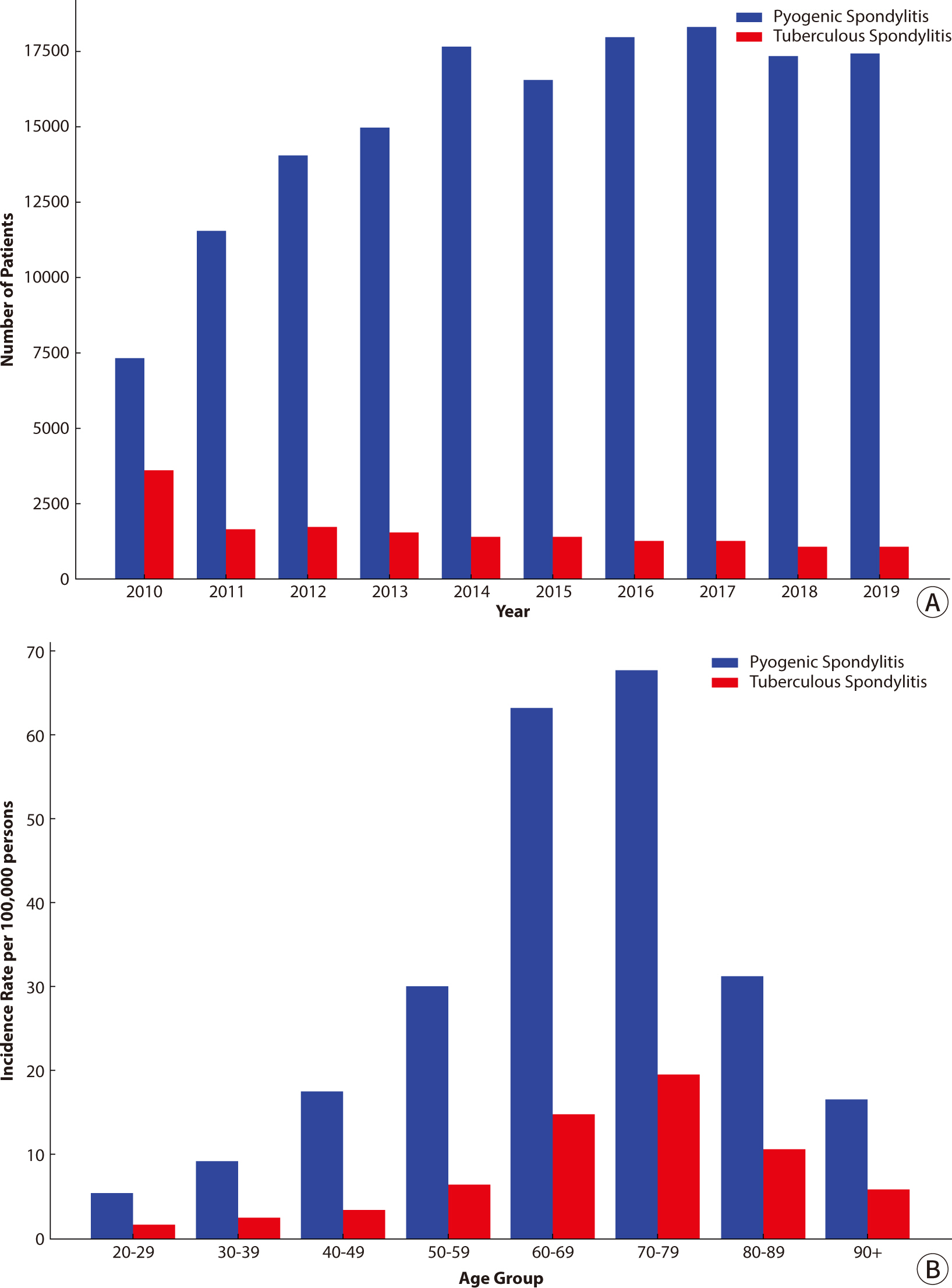
Fig. 1 depicts the incidence of infectious
spondylitis, based on national health insurance data from Korea. A nationwide cohort
study conducted from 2007 to 2016 identified 9,655 cases of the condition [5]. The findings showed an increase in the
number of pyogenic spondylitis cases, risking from 2,431 in 2007 to 4,874 in 2016.
Conversely, the incidence of tuberculous spondylitis declined from 1,756 cases to
594 over the same timeframe. These patterns indicate a shift toward bacterial
infection as the predominant cause of infectious spondylitis in Korea.
Fig. 1.
Incidence of infectious spondylitis in Korea. (A) The number of
infectious spondylitis cases recorded between 2010 and 2019 was determined
using data provided by the Health Insurance Review and Assessment Service
(HIRA) [6]. (B) The incidence rates of
pyogenic spondylitis and tuberculous spondylitis were compared from 2007 to
2016 using data from the Korean National Health Insurance Service (NHIS)
[5].
A more recent study covering the period from 2010 to 2019 further confirmed this
upward trend (Fig. 1A) [6]. Among 169,244 patients, the number of cases increased from
10,991 in 2010 to 18,533 in 2019. In turn, the incidence rate per 100,000 people
climbed from 22.90 to 35.79. This increase is attributed to the aging population,
higher prevalence of chronic diseases, increased use of immunosuppressive therapies,
and greater frequency of invasive spinal procedures [7–9].
Both pyogenic and tuberculous spondylitis exhibited the highest prevalence in
individuals aged 60−79 years (Fig. 1B)
[5]. Interestingly, female patients
predominated in both groups, which contrasts with some international studies
reporting a higher incidence in male patients.
Anatomically, infectious spondylitis predominantly affects the lumbar region,
followed by the thoracic and cervical spine, with the latter comprising less than
10% of cases [10]. Pyogenic spondylitis
primarily targets the lumbar spine, whereas tuberculous spondylitis more commonly
occurs in the thoracic spine, with the lumbar region representing the second most
frequent site [3].
Etiologic microorganisms
In Korea, fungal spondylitis, non-tuberculous mycobacteria, and
Brucella spondylitis are uncommon [9,11,12]. Microorganisms that cause pyogenic
spondylitis typically reach the vertebrae through arterial spread, during spinal
surgery or other procedures, or directly from adjacent sites. Staphylococcus
aureus is the predominant causative agent in pyogenic spondylitis,
followed by Streptococcus species. Gram-negative bacilli are
responsible for 7% to 33% of cases, with Escherichia coli being the
most common among them [10,13,14].
Coagulase-negative staphylococci are implicated in 30% to 32% of pyogenic
spondylitis cases in patients with a history of spinal surgery or other procedures
[15]. Gram-negative bacilli are more
frequently suspected in female patients or in those with previous or concurrent
urinary tract or intra-abdominal infections [10,14]. Table 1 presents the distribution of microorganisms identified
in cases of spontaneous or postoperative pyogenic spondylitis based on Korean data
[15,16].
Table 1.
Distribution of microorganisms identified in patients with spontaneous or
postoperative pyogenic spondylitis
Tuberculous spondylitis primarily results from venous spread originating in the lungs
or other primary lesions. M. tuberculosis can directly infect the
spine from adjacent organs, including the lungs, kidneys, and gastrointestinal
tract. A literature review by Schirmer et al. [17] indicated that the rate of concomitant pulmonary tuberculosis in
patients with tuberculous spondylitis ranges from 8% to 100%. Additionally, a study
from Korea found that 16% of patients with tuberculous spondylitis also had
extrapulmonary tuberculosis, including miliary tuberculosis as well as renal and
lymph node involvement [18].
Comorbidity with other disease
Understanding the distribution of microorganisms based on patient characteristics can
guide clinicians in selecting appropriate empirical antibiotics. An analysis of
Health Insurance Review and Assessment Service data from 2010 to 2019 showed that
patients with infectious spondylitis often exhibit multiple comorbidities. These
include diabetes mellitus (55.1%), rheumatoid arthritis (27.3%), chronic obstructive
pulmonary disease (15.2%), and end-stage renal disease (12.8%) [6]. In a cohort of 586 patients with
culture-proven pyogenic spondylitis, the most common comorbidities were diabetes
(30.7%), solid tumors (14.3%), chronic renal disease (10.4%), and liver cirrhosis
(9.4%) [16]. The study also revealed that
gram-negative infections were relatively prevalent among older patients, women, and
those with cirrhosis or solid tumors. Additionally, methicillin-resistant S.
aureus infection was more frequent in patients with chronic renal
disease than in those without this comorbidity [16].
While one report indicated that diabetes was reported in 17% of 94 patients with
tuberculous spondylitis, no other comorbidities were specifically associated with
this condition [3]. In 2020, Korea had the
highest incidence of tuberculosis among Organisation for Economic Co-operation and
Development countries, with 49 cases per 100,000 population, and an increasing
proportion of new cases were seen in individuals aged 65 and older [19]. Consequently, the range of comorbid
diseases in patients with tuberculous spondylitis may be diverse.
Diagnostic approaches
Clinicians should consider infectious spondylitis in patients presenting with new or
worsening back or neck pain. The onset of symptoms is often gradual and subtle, with
pain typically worsening during weight-bearing activities and subsiding when the
patient lies down. The pain is usually well-localized and can be reproduced through
palpation or percussion over the affected area. Pyogenic spondylitis is relatively
likely among patients who experience back or neck pain along with fever, bloodstream
infection, or infective endocarditis [20].
This condition should also be suspected in patients presenting with fever and new
peripheral neurologic symptoms, with or without back pain. Radiculopathy, which may
manifest as leg pain or weakness, can occur due to nerve root compression or
irritation. In cases involving the thoracic spine, patients often describe a
“belt-like” pain across the chest wall or abdomen, which can be
mistakenly attributed to gastrointestinal, cardiac, or pulmonary conditions.
The initial evaluation of patients with suspected infectious spondylitis should begin
with a comprehensive history and physical examination, including a detailed
neurological assessment. Patients should be asked about any comorbidities, ongoing
infections, and predisposing factors, such as existing non-spinal infections, the
presence of indwelling devices, recent application of surgical instruments, and
spinal injections [10,15]. Initial diagnostic tests include inflammatory markers (WBC
count, ESR, and CRP level), as well as two sets of blood cultures. Spinal imaging is
also critical, with MRI being the preferred method when available. Additionally,
plain X-rays, including anteroposterior and lateral views, along with
flexion/extension views, should be obtained for baseline evaluation in all cases
[21]. However, native X-rays exhibit low
specificity for diagnosing infectious spondylitis, with these examinations primarily
detecting advanced cases characterized by vertebral endplate irregularities or a
reduction in intervertebral disc height.
MRI with intravenous gadolinium contrast is the preferred imaging method due to its
increased sensitivity and specificity. It offers superior visualization of potential
infection spread to the epidural and paravertebral spaces [22]. Clinicians should obtain T2-weighted and post-contrast
T1-weighted images with fat suppression. Typical MRI findings indicative of
infection include abnormal signals from the intervertebral discs, destruction of the
vertebral body endplates adjacent to the disc, and bone marrow edema. However, these
findings can also be present in non-infectious spinal conditions, necessitating
collaboration between clinicians and radiologists to achieve accurate diagnosis and
differentiation [22].
For patients unable to undergo MRI, CT can be used to assess the osseous anatomy and,
with the addition of contrast, can also reveal involvement of paraspinal and
epidural soft tissues. Recent studies have suggested that fluoro-2-deoxyglucose
PET/CT may represent a complementary tool to MRI for differentiating between
tuberculous and pyogenic spondylitis [23], as
well as for assessing disease activity [23,24]. PET/CT offers superior
spatial resolution and improved detection of metastatic infection. The combination
of positive blood cultures, imaging findings, and clinical symptoms can often
confirm a diagnosis of infectious spondylitis [25]. In blood cultures of patients suspected of having pyogenic
spondylitis, when microbial growth is present, the necessity of tissue biopsy
remains a topic of debate [20]. A Korean
retrospective study involving 141 patients with pyogenic spondylitis, who exhibited
positive blood and tissue cultures, reported a 95.7% concordance rate in bacterial
identification [26]. Discordant results were
typically characterized by the growth of a single species in one sample and multiple
species, including the initially identified species, in the other. These findings
suggest that in cases with positive blood cultures, tissue biopsy may not be
necessary for the microbiological diagnosis of pyogenic spondylitis (Fig. 2).
Fig. 2.
Approach to diagnosing a patient with suspected infectious
spondylitis.
Inflammatory markers such as WBC count, CRP level, and ESR are typically elevated in
acute infections but may be normal in chronic cases [26]. Kim et al. [3] found that
patients with pyogenic spondylitis exhibited significantly higher levels of ESR and
CRP compared to those with tuberculous spondylitis. Notably, tuberculosis infection
rates remain high among elderly Koreans, warranting careful consideration in this
demographic [18,27]. Tuberculous spondylitis should be suspected in cases
involving slow disease progression over several months or when extraspinal
tuberculosis is detected [3,18]. Diagnosis is confirmed through tissue
biopsy, with mycobacterial culture positivity rates ranging from 69.0% to 85.3%
[28]. Polymerase chain reaction
techniques have been employed for the rapid identification of mycobacteria in
formaldehyde-fixed, paraffin-embedded tissue specimens. Tissue biopsy is also
indicated when blood cultures fail to establish a microbiologic diagnosis for
pyogenic spondylitis. The two most widely recognized methods are image-guided
percutaneous needle biopsy and open biopsy. Once tissue biopsy is performed,
specimens should be sent for both microbiologic and histopathologic examination.
Needle biopsy specimens can be obtained percutaneously through CT or
fluoroscopically guided biopsy, with diagnostic yields of 44% and 55%, respectively
[29]. If needle biopsy is indicated for
patients with concurrent paraspinal inflammation or abscess, samples should be
collected from paraspinal rather than spinal tissues [30]. Open surgical biopsy is considered the most reliable
method, with a 76% diagnostic yield according to a recent systematic review [31]; however, the impact of prior antibiotic
use requires further clarification. Some experts suggest that in patients with
pyogenic spondylitis who have been exposed to antibiotics but show no signs of
sepsis or severe sepsis, a certain interval should elapse before biopsy is performed
[32].
A second percutaneous biopsy may be warranted if the initial biopsy does not yield a
diagnosis, although the precise benefit of this procedure is still uncertain [33]. It is advisable to wait at least 3 days
after the initial biopsy before repeating the procedure, by which time most positive
cultures from the first biopsy should have been obtained [34]. Alternatively, if the first image-guided biopsy yields a
negative result, proceeding with an open biopsy as the next step is reasonable
(Fig. 2).
If the microbial etiology is not identified, empiric treatment becomes necessary.
Empiric antibiotics should be promptly administered to critically ill patients
showing signs of sepsis or those being taken to the operating room for neurologic
compromise. The initiation of empiric treatment should be based on the most likely
microbial etiology. To select the appropriate empiric antibiotics for a patient with
pyogenic spondylitis of unknown microbial etiology, factors such as medical history,
demographic characteristics, clinical features, and imaging results must be
considered [4,16]. If the patient has not undergone spinal surgery, vancomycin need
not be included in the empiric antibiotic regimen due to the low risk of
methicillin-resistant S. aureus or methicillin-resistant
coagulase-negative staphylococci [13,15,35].
A first-generation cephalosporin is suitable for the treatment of suspected
community-acquired pyogenic spondylitis. Alternative options include a
fluoroquinolone with rifampin, or a fluoroquinolone plus a
beta-lactam/beta-lactamase inhibitor [36,37]. If the patient has
exhibited previous or concurrent urinary tract infection or intra-abdominal
infection, empiric antibiotics should provide coverage for gram-negative bacilli
[10]. Therapy should be adjusted
according to bacteriologic test results. Most cases of pyogenic spondylitis are
treated conservatively, with favorable outcomes. A recent study has established that
a 6-week course of systemic antibiotics is sufficient for most cases [38]. However, a longer duration of therapy may
be required in certain situations, such as infections with extensive spread to
paraspinal soft tissues, undrained paravertebral abscesses, or extensive bone
destruction. Transitioning to oral antibiotics with high bioavailability is
considered acceptable.
In cases of culture-negative infectious spondylitis, which typically involve
long-term and broad-spectrum antibiotic treatment, this strategy can result in
avoidable side effects and contribute to antibiotic resistance. One prior report
indicated favorable outcomes with the use of cefazolin in hematogenous pyogenic
spondylitis and with vancomycin in post-procedural pyogenic spondylitis among
patients with culture-negative pyogenic spondylitis [39].
Treatment
The most severe complication of infectious spondylitis is neurologic impairment,
which can occur secondary to either abscess formation or bony collapse. Treatment
objectives include saving the patient’s life, alleviating pain, preventing or
reversing neurologic deficits, eradicating the infection, and restoring spinal
stability. To meet these treatment objectives, management principles encompass: (1)
establishing an accurate microbiological diagnosis; (2) administering appropriate
antimicrobials; (3) immobilizing the spine; and (4) carefully monitoring for
clinical and radiographic evidence of spinal instability, as well as for progression
of the infection or neurological deterioration.
The treatment regimens for tuberculous spondylitis align with those for pulmonary
tuberculosis. For most patients receiving rifampin for susceptible tuberculosis, a
6- to 9-month course of therapy is sufficient [40]. To date, no formal data are available on the efficacy of newer
drugs in the treatment of osteoarticular tuberculosis.
While receiving antimicrobial therapy, patients should be carefully monitored for
clinical signs of soft tissue extension or abscess, as well as for symptoms of cord
compression. Additionally, clinicians should track inflammatory markers,
specifically ESR and CRP levels, with weekly assessments [20]. CRP levels tend to normalize more quickly than ESR
following successful treatment or after uncomplicated spinal fusion surgery [41]. Routine anteroposterior and lateral
radiographs centered on the affected disc are recommended at 1 and 3 months into
antimicrobial therapy, and again 3 months after the cessation of treatment [42]. For the cervical or lumbar spine,
orthopedic surgeons advise obtaining follow-up flexion/extension films to reliably
detect potential instability or to confirm bone fusion. In patients who are
clinically improving while on treatment, routine follow-up MRI is unnecessary, as
imaging findings may not correspond with clinical progress [43].
Surgical intervention, which may include procedures such as incision and drainage,
decompression, corpectomy, and fusion, is sometimes required. Patients presenting
with neurological deficits such as weakness, paresthesia, and urinary retention, as
well as those with radiographic signs of epidural or paravertebral abscess or actual
or impending spinal cord compression, should be evaluated for surgical
decompression. Interventional radiology has become increasingly important in
managing psoas muscle abscesses. Continuous monitoring for the development or
progression of neurological signs is crucial, yet it is frequently overlooked.
Epidural abscesses can lead to abrupt neurological deficits. A spinal epidural
abscess, a potentially severe complication of infectious spondylitis, can spread
through septic thrombosis of the epidural veins. Since skip lesions, or
noncontiguous abscesses, may occur in 15% of overall cases [44], imaging of the entire spine is recommended.
Relative indications for surgery include uncertain diagnosis, lack of clinical
improvement following antimicrobial treatment, or significant progressive spinal
deformity accompanied by biomechanical instability. However, guidelines do not offer
a detailed and practical description of surgical interventions for cases of
spondylitis that are resistant to conservative treatment [20]. Decisions regarding surgery should be made in close
consultation with surgeons.
In the early phase of infectious spondylitis, bed rest is recommended until the acute
pain improves. Both bed rest and spinal immobilization are crucial, particularly in
cases of vertebral destruction. Once the acute pain has subsided, ambulation with an
appropriate brace is advised. Patients with thoracic infections should use a
thoracolumbar sacral orthosis, while those with lumbosacral infections are advised
to use a lumbar sacral orthosis. The duration of thoracolumbar sacral orthosis brace
usage varies depending on factors such as the patient’s response to
treatment, the nature of the infection, and the overall health and stability of the
spine. Research indicates that approximately 30% of patients may experience a
progression of deformity during the first 6 to 8 weeks [45]. Typically, patients may need to wear the brace
continuously for several weeks to months, with the duration of use gradually
decreasing as healing progresses. Patients should be monitored throughout the
treatment and for 1 year after its completion to detect any relapses [46].
Prognosis
Most patients experience a gradual improvement in back pain after the initiation of
treatment, with the pain typically resolving after bone fusion occurs. However,
clinicians must communicate to patients and their caregivers that back pain may
persist. A systematic review of the clinical characteristics of infectious
spondylitis reported an attributable mortality rate of 6% [47]. The functional outcome is worse in cases with neurological
deficits, which have been noted in 32% of patients. Additionally, 27% of patients
experience complications that significantly impact their quality of life [47]. In a large retrospective study from Japan,
which included over 7,000 patients with infectious spondylitis, the in-hospital
mortality rate was 6% [48]. Comorbidities
such as diabetes, end-stage kidney disease, cirrhosis, malignancy, and infective
endocarditis were determinants of this mortality rate. Similarly, a retrospective
study from a single center in Korea, which included 116 patients with infectious
spondylitis, reported an in-hospital mortality rate of 6% and a relapse rate of 8%
[9]. Recurrences typically occur within 6
months, and rarely up to 1 year, after the completion of antibiotic therapy [35].
Conclusion
Infectious spondylitis is a serious condition that necessitates timely diagnosis and
effective treatment to reduce the risk of complications, such as neurological
impairment. The incidence of pyogenic spondylitis has risen in Korea, while
tuberculous spondylitis remains a key concern due to the persistent prevalence of
tuberculosis. Accurate microbiological diagnosis, appropriate antimicrobial therapy,
and vigilant monitoring are essential for the management of infectious spondylitis.
Both medical and surgical interventions are important and are chosen based on the
severity and progression of the disease. Clinicians must recognize the variety of
etiological microorganisms, consider patient comorbidities, and understand the vital
role of a multidisciplinary approach in delivering optimal care. Ongoing education
and research are imperative to establish standardized treatment protocols and
improve prognoses for patients with infectious spondylitis.
Authors' contributions
All work was done by Kyung-Hwa Park.
Conflict of interest
No potential conflict of interest relevant to this article was reported.
Funding
This work was supported by the Chonnam National University Hospital Biomedical
Research Institute (BCR124055). The funders had no role in the study design,
data collection and analysis, decision to publish, or preparation of the
manuscript.
3. Kim CJ, Song KH, Jeon JH, Park WB, Park SW, Kim HB, et al. A comparative study of pyogenic and tuberculous
spondylodiscitis. Spine 2010;35(21):E1096-E1100.
4. Lee Y, Kim BJ, Kim SH, Lee SH, Kim WH, Jin SW. Comparative analysis of spontaneous infectious spondylitis:
pyogenic versus tuberculous. J Korean Neurosurg Soc 2018;61(1):81-88.
5. Kim YJ, Hong JB, Kim YS, Yi J, Choi JM, Sohn S. Change of pyogenic and tuberculous spondylitis between 2007 and
2016 year: a nationwide study. J Korean Neurosurg Soc 2020;63(6):784-793.
6. Son HJ, Kim M, Kim DH, Kang CN. Incidence and treatment trends of infectious spondylodiscitis in
South Korea: a nationwide population-based study. PLoS One 2023;18(6):e0287846.
7. Moon YJ, Kim DY, Song KJ, Kim YJ, Lee KB. Relationship between percutaneous procedures and lumbar
infections based on data from The National Health Insurance Review &
Assessment Service of Korea. Clin Spine Surg 2016;29(1):E55-E60.
8. Noh SH, Zhang HY, Lee SH, Choi JK, Chin DK. Changes in trends of spondylitis in Korea based on a nationwide
database. Yonsei Med J 2019;60(5):487-489.
9. Kim YI, Kim SE, Jang HC, Jung SI, Song SK, Park KH. Analysis of the clinical characteristics and prognostic factors
of infectious spondylitis. Infect Chemother 2011;43(1):48-54.
10. Kang SJ, Jang HC, Jung SI, Choe PG, Park WB, Kim CJ, et al. Clinical characteristics and risk factors of pyogenic spondylitis
caused by gram-negative bacteria. PLoS One 2015;10(5):e0127126.
11. Kim CJ, Kim UJ, Kim HB, Park SW, Oh M, Park KH, et al. Vertebral osteomyelitis caused by non-tuberculous mycobacteria:
predisposing conditions and clinical characteristics of six cases and a
review of 63 cases in the literature. Infect Dis 2016;48(7):509-516.
12. Lee JY, Jeon Y, Ahn MY, Ann HW, Jung IY, Jung W, et al. An imported case of Brucella melitensis infection in South
Korea. Infect Chemother 2018;50(2):149-152.
14. Park KH, Cho OH, Jung M, Suk KS, Lee JH, Park JS, et al. Clinical characteristics and outcomes of hematogenous vertebral
osteomyelitis caused by gram-negative bacteria. J Infect 2014;69(1):42-50.
15. Kim UJ, Bae JY, Kim SE, Kim CJ, Kang SJ, Jang HC, et al. Comparison of pyogenic postoperative and native vertebral
osteomyelitis. Spine J 2019;19(5):880-887.
16. Kim DY, Kim UJ, Yu Y, Kim SE, Kang SJ, Jun KI, et al. Microbial etiology of pyogenic vertebral osteomyelitis according
to patient characteristics. Open Forum Infect Dis 2020;7(6):ofaa176
18. Kim CJ, Kim EJ, Song KH, Choe PG, Park WB, Bang JH, et al. Comparison of characteristics of culture-negative pyogenic
spondylitis and tuberculous spondylitis: a retrospective
study. BMC Infect Dis 2016;16(1):560
20. Berbari EF, Kanj SS, Kowalski TJ, Darouiche RO, Widmer AF, Schmitt SK, et al. 2015 Infectious Diseases Society of America (IDSA) clinical
practice guidelines for the diagnosis and treatment of native vertebral
osteomyelitis in adults. Clin Infect Dis 2015;61(6):e26-e46.
21. Lener S, Hartmann S, Barbagallo GMV, Certo F, Thomé C, Tschugg A. Management of spinal infection: a review of the
literature. Acta Neurochir 2018;160(3):487-496.
22. Ryu S, Kim YJ, Lee S, Ryu J, Park S, Hong JU. Pathophysiology and MRI findings of infectious spondylitis and
the differential diagnosis. J Korean Soc Radiol 2021;82(6):1413-1440.
23. Lee IS, Lee JS, Kim SJ, Jun S, Suh KT. Fluorine-18-fluorodeoxyglucose positron emission
tomography/computed tomography imaging in pyogenic and tuberculous
spondylitis: preliminary study. J Comput Assist Tomogr 2009;33(4):587-592.
24. Jeon I, Kong E. Application of simultaneous 18F-FDG PET/MRI for evaluating
residual lesion in pyogenic spine infection: a case report. Infect Chemother 2020;52(4):626-633.
25. Bae JY, Kim CJ, Kim UJ, Song KH, Kim ES, Kang SJ, et al. Concordance of results of blood and tissue cultures from patients
with pyogenic spondylitis: a retrospective cohort study. Clin Microbiol Infect 2018;24(3):279-282.
26. Herren C, Jung N, Pishnamaz M, Breuninger M, Siewe J, Sobottke R. Spondylodiscitis: diagnosis and treatment options. Dtsch Arztebl Int 2017;114(51-52):875-882.
28. Lee CM, Lee Y, Kang SJ, Kang CK, Choe PG, Song KH, et al. Positivity rates of mycobacterial culture in patients with
tuberculous spondylitis according to methods and sites of biopsies: an
analysis of 206 cases. Int J Infect Dis 2022;121:161-165.
29. McNamara AL, Dickerson EC, Gomez-Hassan DM, Cinti SK, Srinivasan A. Yield of image-guided needle biopsy for infectious discitis: a
systematic review and meta-analysis. AJNR Am J Neuroradiol 2017;38(10):2021-2027.
30. Kim CJ, Kang SJ, Choe PG, Park WB, Jang HC, Jung SI, et al. Which tissues are best for microbiological diagnosis in patients
with pyogenic vertebral osteomyelitis undergoing needle
biopsy? Clin Microbiol Infect 2015;21(10):931-935.
34. Yeh KJ, Husseini JS, Hemke R, Nelson SB, Chang CY. CT-guided discitis-osteomyelitis biopsies with negative
microbiology: how many days should we wait before repeating the
biopsy? Skeletal Radiol 2020;49(4):619-623.
35. Lee YD, Jeon YH, Kim YH, Ha KY, Hur JW, Ryu KS, et al. Clinical characteristics and outcomes of patients with
culture-negative pyogenic spondylitis according to empiric glycopeptide
use. Infect Chemother 2019;51(3):274-283.
36. Park KH, Kim DY, Lee YM, Lee MS, Kang KC, Lee JH, et al. Selection of an appropriate empiric antibiotic regimen in
hematogenous vertebral osteomyelitis. PLoS One 2019;14(2):e0211888.
37. The Korean Society for Chemotherapy, The Korean Society of
Infectious Diseases, The Korean Orthopaedic
Association. Clinical guidelines for the antimicrobial treatment of bone and
joint infections in Korea. Infect Chemother 2014;46(2):125-138.
38. Bernard L, Dinh A, Ghout I, Simo D, Zeller V, Issartel B, et al. Antibiotic treatment for 6 weeks versus 12 weeks in patients with
pyogenic vertebral osteomyelitis: an open-label, non-inferiority,
randomised, controlled trial. Lancet 2015;385(9971):875-882.
39. Yoon SH, Chung SK, Kim KJ, Kim HJ, Jin YJ, Kim HB. Pyogenic vertebral osteomyelitis: identification of microorganism
and laboratory markers used to predict clinical outcome. Eur Spine J 2010;19(4):575-582.
40. Pandita A, Madhuripan N, Pandita S, Hurtado RM. Challenges and controversies in the treatment of spinal
tuberculosis. J Clin Tuberc Other Mycobact Dis 2020;19:100151
42. Grados F, Lescure FX, Senneville E, Flipo RM, Schmit JL, Fardellone P. Suggestions for managing pyogenic (non-tuberculous) discitis in
adults. Joint Bone Spine 2007;74(2):133-139.
47. Mylona E, Samarkos M, Kakalou E, Fanourgiakis P, Skoutelis A. Pyogenic vertebral osteomyelitis: a systematic review of clinical
characteristics. Semin Arthritis Rheum 2009;39(1):10-17.
48. Akiyama T, Chikuda H, Yasunaga H, Horiguchi H, Fushimi K, Saita K. Incidence and risk factors for mortality of vertebral
osteomyelitis: a retrospective analysis using the Japanese diagnosis
procedure combination database. BMJ Open 2013;3(3):e002412.
Infectious Spondylitis Caused by Staphylococcus lugdunensis Following an L-PEN Procedure: A Case Report Youngkwon Yang, Kyungryeol Kang, Eundong Lee, Jeongeun Lee, Seeun Jung Cureus.2026;[Epub] CrossRef
Percutaneous Endoscopic Debridement and Drainage for Infectious Spondylodiscitis Under Local Anesthesia Ji-Ho Jung, Jong-Hoon Jeong, Jong-Hwan Hong, Moon-Soo Han, Jung-Kil Lee Journal of Minimally Invasive Spine Surgery and Technique.2026; 11(1): 163. CrossRef
Global, regional, and national burden of bone and joint infections, 1990–2021: a comprehensive analysis of trends, pathogens, and antimicrobial resistance Ke Xu, Xia Zhao, Shuangqing Zhang Frontiers in Cellular and Infection Microbiology.2026;[Epub] CrossRef
Pyogenic Spondylitis Due to Erysipelothrix rhusiopathiae Infection: A Case Report Yusuke Oshita, Takeshi Eguro, Satoshi Kimura, Keikichi Kawasaki, Yoshifumi Kudo Cureus.2025;[Epub] CrossRef
Epidemiology and management of infectious spondylitis in Korea: a
narrative review
Fig. 1.
Incidence of infectious spondylitis in Korea. (A) The number of
infectious spondylitis cases recorded between 2010 and 2019 was determined
using data provided by the Health Insurance Review and Assessment Service
(HIRA) [6]. (B) The incidence rates of
pyogenic spondylitis and tuberculous spondylitis were compared from 2007 to
2016 using data from the Korean National Health Insurance Service (NHIS)
[5].
Fig. 2.
Approach to diagnosing a patient with suspected infectious
spondylitis.
Fig. 1.
Fig. 2.
Epidemiology and management of infectious spondylitis in Korea: a
narrative review
Distribution of microorganisms identified in patients with spontaneous or
postoperative pyogenic spondylitis