1Department of Pathology, Seoul National University Hospital, Seoul National University College of Medicine, Seoul, Korea
*Corresponding author: Haeryoung Kim,
Department of Pathology, Seoul National University Hospital, Seoul National
University College of Medicine, 103 Daehak-no, Jongno-gu, Seoul 03080, Korea,
E-mail: haeryoung.kim@snu.ac.kr
• Received: August 19, 2024 • Accepted: September 26, 2024
This is an Open-Access article distributed under the terms of the
Creative Commons Attribution Non-Commercial License (http://creativecommons.org/licenses/by-nc/4.0) which permits
unrestricted non-commercial use, distribution, and reproduction in any
medium, provided the original work is properly cited.
Hepatocellular carcinoma (HCC) remains a leading cause of cancer-related deaths
worldwide, with poor clinical outcomes due to challenges in early detection and
limited efficacy of current treatments such as receptor tyrosine kinase
inhibitors and immunotherapy. HCC exhibits significant heterogeneity at both
histopathological and molecular levels, complicating its management but offering
potential for personalized therapeutic approaches. This review outlines the
morpho-molecular heterogeneity of HCC and summarizes various histological
subtypes, including steatohepatitic, clear cell, macrotrabecular-massive,
scirrhous, lymphocyte-rich, and fibrolamellar HCCs. Each subtype possesses
distinct clinical, histological, and molecular features; for instance,
steatohepatitic HCC is associated with metabolic dysfunction and shows
IL-6/JAK/STAT activation, while clear cell HCCs often have IDH1
mutations and favorable prognosis. The macrotrabecular-massive subtype is linked
to poor outcomes and TP53 mutations, whereas scirrhous HCCs
express stemness markers and have TSC1/TSC2 mutations.
Lymphocyte-rich HCCs are characterized by immune cell infiltration and better
prognosis. CTNNB1-mutated HCCs show specific morphological
features and may benefit from targeted therapies. Understanding these subtypes
and associated molecular alterations is crucial for developing effective
diagnostic and therapeutic strategies, including potential predictive biomarkers
and personalized treatments. Additionally, the identification of patterns like
vessels-encapsulating-tumor-clusters offers prognostic implications and may
guide therapeutic decisions. Recent molecular studies have enhanced our
comprehension of HCC heterogeneity, laying the groundwork for more personalized
approaches. Pathologists play a vital role in recognizing these subtypes, aiding
in prognosis prediction and treatment planning. Advances in digital pathology
and artificial intelligence may further facilitate biomarker research,
ultimately improving patient outcomes in HCC management.
Hepatocellular carcinoma (HCC) is the most common primary liver cancer, accounting
for 75%–80% of primary liver malignancies [1]. HCC mostly develops in the background of chronic liver disease, the
most common etiologies being HBV or HCV infection, and chronic alcohol abuse. During
the recent year, metabolic syndrome has become another major risk factor for HCC,
even in Asian countries [2,3].
The clinical outcome of patients with HCC is generally poor, mainly due to
difficulties in early detection and limited treatment options for advanced disease
[4,5]. Currently, systemic therapy, such as receptor tyrosine kinase inhibitors
and immunotherapy, is the treatment of choice for the patients with unresectable HCC
[5]. However, the efficacy of systemic
therapy in HCC is still unsatisfactory with survival benefit of 1–3 months,
objective response rate less than 30%, and a high incidence of adverse events [6,7]. In
this context, understanding the heterogeneity of HCC will play a key role in
developing effective diagnostic and therapeutic strategies, by offering potential
predictive biomarkers and personalized approaches for HCC management.
Ethics statement
It is a literature database-based review; therefore, neither approval by the
institutional review board nor obtainment of informed consent was required.
Morpho-molecular heterogeneity of hepatocellular carcinoma
HCC is typically composed of tumor cells showing hepatocytic differentiation with
variable degrees of atypia [8]. While HCC
recapitulates the cytoarchitectural morphology of the liver to varying extents,
there are alterations in the hepatic microarchitecture such as loss of portal
tracts, reduction or loss of the reticulin framework, and thickening of the hepatic
plates. Neoangiogenesis occurs in HCCs, in the form of sinusoidal capillarization
and unpaired arteries [8]. These changes also
result in the characteristic imaging features, such as the early enhancement on
contrast enhanced CT/MRI.
However, there is considerable heterogeneity of HCC, in the microscopic growth
patterns, cytological features, and histological grade. About 50% of HCCs
demonstrate mixed patterns of growth: trabecular, pseudoglandular, solid, and
macrotrabecular (Fig. 1) [9]. In addition, while most HCCs demonstrate cytological
features that recapitulate those of normal hepatocytes (i.e. polygonal cells with
abundant eosinophilic cytoplasm), some HCCs show extensive areas with clear cell
change, fatty change, and cholestasis. Cytoplasmic inclusions (e.g., hyaline bodies,
Mallory-Denk bodies, and pale bodies) may also be seen in some tumor cells [8]. Histological grading is currently performed
according to either the four-tiered modified Edmondson and Steiner system or the
three-tiered World Health Organization (WHO) grading system [9–11].
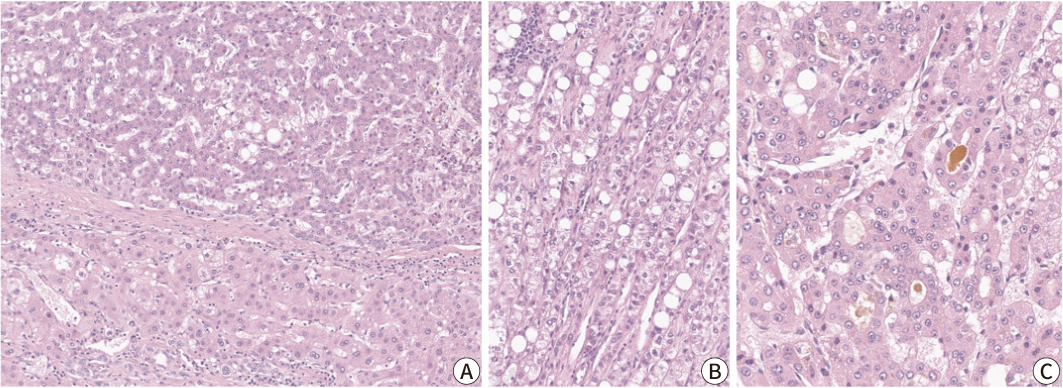
Fig. 1.
Microscopic features of a typical hepatocellular carcinoma (HCC). (A)
Non-neoplastic hepatocytes (lower half) and HCC tumor cells (upper half) are
separated by a fibrous capsule. (B) Trabecular pattern HCC with steatosis.
(C) Pseudoglandular pattern HCC with cholestasis (Hematoxylin-Eosin stain,
original magnification ×100 [A,B], ×200 [C]).
Recent advances in genomic techniques have unraveled the heterogeneity in the
mutational landscape of HCC [12,13]. The most frequently mutated genes include
TERT promoter, TP53, CTNNB1, ARID1A, ARID2, JAK1, ALB,
AXIN1, NFE2L2, and RPS6KA3 [12]. In addition, gene expression profiling studies have
suggested several molecular subclasses of HCC that correlate with the
clinicopathological features, providing the foundation for an integrated
morphological-molecular classification of HCC [14]. In the past two decades, there have been many efforts to establish
a subclassification system that better categorizes HCCs with distinct clinical,
histological, and molecular features (Table
1) [13,15–19]. HCC can be
subclassified into two major groups, the proliferative class and the
non-proliferative class. The proliferative class is characterized by high
chromosomal instability and TP53 mutations, and is associated with
poor histological differentiation, frequent vascular invasion, increased
alpha-fetoprotein (AFP) level, and overall poor clinical outcome [16,18].
On the other hand, the non-proliferative class displays chromosomal stability and a
well-differentiated phenotype with less frequent vascular invasion [16,18].
CTNNB1-mutated HCCs belong to the latter group: these
demonstrate frequent cholestasis and less immune cell infiltration on histology
[14,20].
Table 1.
Integrated morphological-molecular classification of hepatocellular
carcinoma
Currently, approximately 35% of HCC can be further subclassified into histological
subtypes with distinct morphological, clinicopathological and molecular
characteristics [9]. The following section
will summarize the clinicopathological and molecular features of these different
subtypes.
Steatohepatitic hepatocellular carcinoma
The steatohepatitic subtype of HCC demonstrates the key histological features of
non-neoplastic steatohepatitis, including steatosis, pericellular fibrosis, cell
ballooning, inflammation, and Mallory-Denk bodies, and these features occupy a major
portion (>50%) of the tumor (Fig. 2)
[21]. This subtype has been more
frequently identified in patients with underlying metabolic dysfunction-associated
steatotic liver disease and alcohol abuse, and its relative frequency has been
reported to be between 5% and 20% [3,21]. Steatohepatitic HCC has been associated
with less frequent vascular invasion and satellite nodules; however, its prognosis
appears to be similar to that of conventional HCC [14]. Key molecular alterations associated with steatohepatitic HCC
include IL-6/JAK/STAT activation, while CTNNB1, TERT promoter and
TP53 mutations have been found to be less frequent in these
tumors [14].
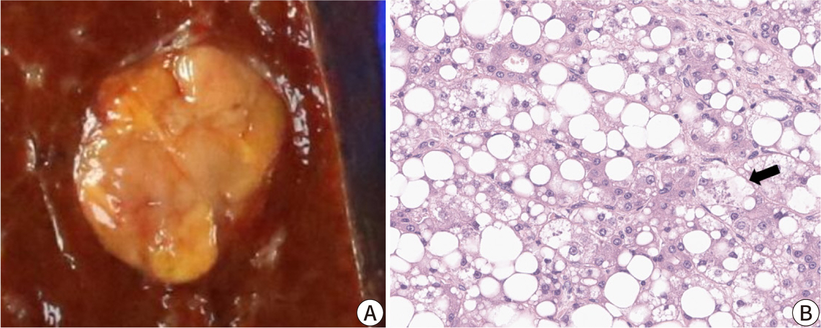
Fig. 2.
Steatohepatitic hepatocellular carcinoma (HCC). (A) The tumor
demonstrates a yellow hue on macroscopy reflecting the lipid component. (B)
High power magnification showing the diffuse steatosis of tumor cells, tumor
cell ballooning (arrow), some inflammatory cells, and pericellular fibrosis
(Hematoxylin-Eosin stain, original magnification ×400).
Clear cell hepatocellular carcinoma
By definition, in clear cell HCCs, more than 80% of tumor cells demonstrate abundant
clear cytoplasm (Fig. 3). The clear cytoplasm
is a result of glycogen accumulation; however, some tumor cells may appear clear due
to lipid droplets, and some degree of steatosis is acceptable for this diagnosis
[22]. The relative frequency of clear
cell HCC has been estimated to be around 3%–7%. Clear cell HCCs are generally
well-differentiated tumors with a favorable prognosis [23]. One study has reported that clear cell HCCs demonstrate
higher frequency of IDH1 mutation, although this mutation is not
sufficient to define the subtype [24].
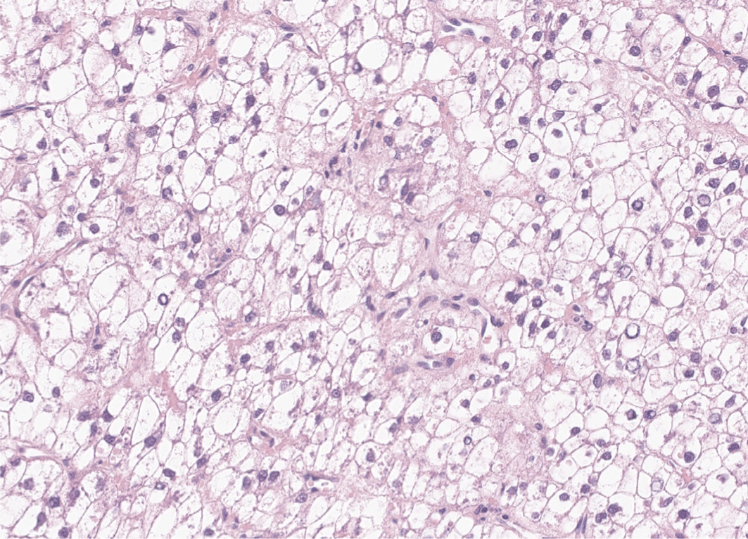
Fig. 3.
Clear cell hepatocellular carcinoma (HCC). Most of the tumor cells
demonstrate clear cytoplasm due to glycogen accumulation (Hematoxylin-Eosin
stain, original magnification ×200).
Macrotrabecular-massive hepatocellular carcinoma
The macrotrabecular-massive subtype of HCC is an HCC in which more than 50% of the
tumor cells assume a macrotrabecular growth pattern, defined as large trabeculae
that are more than 6–10 cells thick (Fig.
4) [25]. This subtype accounts for
approximately 5% of all HCCs and has been strongly associated with elevated serum
AFP levels, high-grade cytological atypia, extensive lymphovascular invasion, more
frequent distant metastasis, and a poor prognosis [25,26]. In addition, the
vessels-encapsulating-tumor-clusters (VETC) pattern of neoangiogenesis, which has
been associated with metastatic dissemination of HCC, is often enriched in this
subtype (Fig. 4) [27,28].
TP53 mutations and FGF19 amplifications have
been more frequently identified in the macrotrabecular-massive subtype of HCC [14].
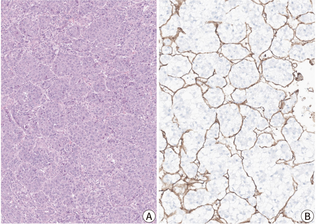
Fig. 4.
Macrotrabecular-massive hepatocellular carcinoma (HCC; A) and
vessels-encapsulating-tumor clusters (VETC) pattern (B). (A)
Macrotrabecular-massive HCC demonstrating thick tumor cell trabeculae, of
more than 10-cell thickness (Hematoxylin-Eosin stain, original magnification
×100). (B) CD34 immunostain highlighting the VETC pattern, where the
CD34-positive endothelial cells completely surround tumor cell
clusters.
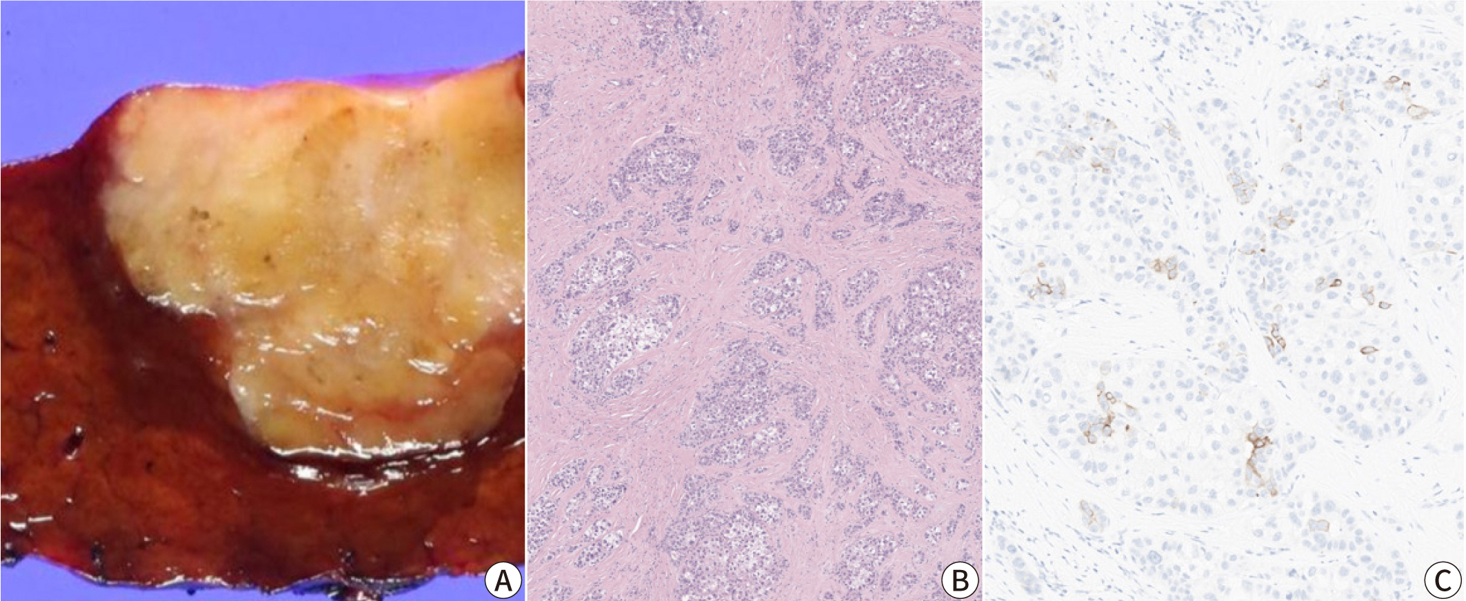
Scirrhous hepatocellular carcinoma
This subtype is characterized by dense intratumoral fibrous stroma (Fig. 5). The scirrhous subtype has a relative
frequency of 4% and often mimics intrahepatic cholangiocarcinoma on imaging [29]. Expression of immunohistochemical markers
associated with stemness (e.g., cytokeratin ([CK]) 7, CK19, and epithelial cell
adhesion molecule) is often seen in scirrhous HCCs, and increased expression of
cholangiocarcinoma-like and stem-cell-like genes have been identified by gene
expression profiling, consistent with the intermediate characteristic of this
subtype [30,31]. Furthermore, scirrhous HCC is associated with frequent
TSC1/TSC2 mutations and transforming growth factor-β
signaling activation [14,30].
Fig. 5.
Scirrhous hepatocellular carcinoma (HCC). (A) The tumor appears as a
firm, yellowish-white and lobulated mass on gross examination, mimicking an
intrahepatic cholangiocarcinoma. (B) Dense intratumoral fibrosis is evident
at low power magnification (Hematoxylin-Eosin stain, original magnification
×40). (C) Immunohistochemical expression of cytokeratin 19 is seen in
a few tumor cells.
Lymphocyte-rich hepatocellular carcinoma
The lymphocyte-rich subtype demonstrates massive intratumoral infiltration of
lymphocytes, which outnumber the tumor cells in most microscopic fields. This
subtype is rare, accounting for less than 1% of all HCCs, but has received much
attention as it has been associated with a favorable clinical outcome [32]. The lymphocyte-rich subtype is associated
with increased programmed death-ligand 1 expression and focal amplification of
chromosome 11q13.3, which is related to the immune checkpoint signature (CD274,
PDCD1, BTLA, CTLA4, HAVCR2, IDO1, and LAG3) [32–35]. Interestingly,
although this subtype is also known as “lymphoepithelioma-like HCC”,
it is not associated with Epstein-Barr virus infection, unlike the
lymphoepithelioma-like tumors arising in other organs, such as the nasopharynx and
stomach [32].
Fibrolamellar hepatocellular carcinoma
Fibrolamellar carcinoma, or fibrolamellar HCC, consists of strands of large
eosinophilic tumor cells with abundant cytoplasm and prominent nucleoli, and
separated by dense intratumoral bands of fibrosis [36]. Fibrolamellar carcinoma accounts for approximately 1% of all HCC,
occurs in younger patients (median age of 25 years), and the background liver is
non-cirrhotic [37]. The prognosis of
fibrolamellar carcinoma appears to be better than that of conventional HCC arising
in cirrhotic livers, but similar to that of HCC in non-cirrhotic livers [37]. DNAJB1-PRKACA gene fusion
has been identified in >95% of cases, and fluorescence in
situ hybridization for PRKACA gene rearrangement is a
useful ancillary test in confirming the diagnosis [38]. Expression of CK7 and CD68 in the tumor cells is another
characteristic of fibrolamellar carcinoma [38].
CTNNB1-mutated hepatocellular carcinoma
CTNNB1 mutations have been reported in approximately 20%–40%
of HCCs [39]. CTNNB1 encodes
β-catenin, which plays a key role in the WNT signaling pathway that regulates
liver function and zonation [40]. In
addition, bile salt transporter expression is dysregulated in these tumors,
histologically manifested by frequent intratumoral cholestasis. Some
CTNNB1-mutated HCCs may be diagnosed by gadoxetic acid-enhanced
MRI, due to the upregulation of the organic anion transporting polypeptide 1B3
(OATP1B3) [41]. Histologically,
CTNNB1-mutated HCCs are typically well-differentiated tumors
with microtrabecular and/or pseudoglandular growth patterns, intratumoral
cholestasis, and less immune cell infiltration compared to
non-CTNNB1-mutated HCCs [14,20]. However,
CTNNB1-mutated HCCs are not morphologically homogeneous, with
approximately 40% not demonstrating the “classic CTNNB1
morphology” [42]. Immunohistochemical
studies for β-catenin (nuclear expression) and glutamine synthetase (diffuse,
strong and homogeneous expression) may serve as useful surrogate markers for
CTNNB1 mutation.
Hepatocellular carcinoma with stemness-related marker expression
HCC with stemness-related marker expression, or progenitor HCC, is defined as HCC
expressing stemness-related markers, e.g., CK19, in >5% of the tumor cells
[15]. This subset of HCCs differ from
combined hepatocellular-cholangiocarcinoma, as they are morphologically compatible
with HCC, and there is no evidence of glandular differentiation or mucin production
in these tumors. They are associated with increased serum AFP levels, frequent
vascular invasion, poor histological differentiation, high recurrence rate,
resistance to systemic chemotherapy and locoregional treatment, and overall poor
prognosis [43]. HCCs with stemness-related
marker expression more frequently demonstrate TP53 mutations and
chromosomal instability, and increased PD-L1 expression [33,34].
Hepatocellular carcinomas with vessels-encapsulating-tumor-clusters
pattern
The VETC phenotype is defined by the presence of VETC pattern in more than 55% of the
tumor area, characterized by CD34-positive vessels that encapsulate and isolate
individual tumor clusters, forming a cobweb-like pattern (Fig. 4) [27,28,44].
The VETC pattern is often found in the macrotrabecular-massive subtype of HCC (7.8%)
and is associated with aggressive behavior and metastatic dissemination [27,28].
It has been reported that VETC pattern is related to a novel mechanism of
metastasis, independent of epithelial-to-mesenchymal transition [44]. Furthermore, patients with VETC-positive
HCC have shown greater survival benefits from sorafenib therapy compared to those
with VETC-negative HCC, suggesting that the VETC pattern may serve as a potential
predictive marker for sorafenib response [28]. Correlation between the VETC pattern on histology and a rim arterial
phase hyperenhancement in arterial phase imaging suggests a role for imaging in the
prognostication of HCC [45].
Other rare histological subtypes of HCC have been described. The chromophobe subtype
of HCC has tumor cells with clear to pale cytoplasm and mainly bland nuclei with
focal areas of striking nuclear atypia [46].
Chromophobe subtype is strongly associated with alternative lengthening of
telomeres, a telomerase-independent mechanism of telomere maintenance, which can be
detected by fluorescence in situ hybridization [46]. Its prognosis is currently known to be
similar to that of conventional HCC [47].
Neutrophil-rich HCC is characterized by marked intratumoral neutrophilic
infiltration, granulocyte colony-stimulating factor production by tumor cells, and a
poor prognosis [47]. The tumor cells are
often poorly differentiated, and focal sarcomatoid differentiation can be observed
[48].
Conclusion
Recent molecular studies have significantly enhanced our understanding of the
morphological and molecular heterogeneity of HCC, providing the foundation for more
effective and personalized treatment strategies. Pathologists are becoming
increasingly aware of the histomorphological heterogeneity of HCC, and the
specification of the various subtypes of HCC has helped pathologists understand the
histology of HCC in more detail and the various differential diagnoses and
diagnostic pitfalls for each variant. The correlation between the histomorphology
and the molecular and biological features suggests the role of histology in the
prediction of therapeutic response and prognosis. This may be further facilitated by
the recent advances in digital pathology and artificial intelligence-based biomarker
research.
Authors' contributions
Project administration: not applicable
Conceptualization: Chung W, Kim H
Methodology & data curation: not applicable
Funding acquisition: Kim H
Writing – original draft: Chung W
Writing – review & editing: Chung W, Kim H
Conflict of interest
No potential conflict of interest relevant to this article was reported.
Funding
This was supported by the National Research Foundation of Korea (NRF) grant
funded by the Korea government (MSIT) (NRF-2022R1A2C2010348).
Data availability
Not applicable.
Acknowledgments
Not applicable.
Supplementary materials
Not applicable.
References
1. Bray F, Ferlay J, Soerjomataram I, Siegel RL, Torre LA, Jemal A. Global cancer statistics 2018: GLOBOCAN estimates of incidence
and mortality worldwide for 36 cancers in 185 countries. CA Cancer J Clin 2018;68(6):394-424.
6. Doycheva I, Thuluvath PJ. Systemic therapy for advanced hepatocellular carcinoma: an update
of a rapidly evolving field. J Clin Exp Hepatol 2019;9(5):588-596.
8. Burt AD, Ferrell LD, Hübscher SG. MacSween's pathology of the liver. Amsterdam: Elsevier Health Sciences; 2022.
9. Nagtegaal ID, Odze RD, Klimstra D, Paradis V, Rugge M, Schirmacher P, et al. The 2019 WHO classification of tumours of the digestive
system. Histopathology 2020;76(2):182-188.
11. Martins-Filho SN, Paiva C, Azevedo RS, Alves VAF. Histological grading of hepatocellular carcinoma: a systematic
review of literature. Front Med 2017;4:193
13. The Cancer Genome Atlas Research Network. Comprehensive and integrative genomic characterization of
hepatocellular carcinoma. Cell 2017;169(7):1327-1341.E23.
14. Calderaro J, Couchy G, Imbeaud S, Amaddeo G, Letouzé E, Blanc JF, et al. Histological subtypes of hepatocellular carcinoma are related to
gene mutations and molecular tumour classification. J Hepatol 2017;67(4):727-738.
15. Lee JS, Heo J, Libbrecht L, Chu IS, Kaposi-Novak P, Calvisi DF, et al. A novel prognostic subtype of human hepatocellular carcinoma
derived from hepatic progenitor cells. Nat Med 2006;12(4):410-416.
16. Boyault S, Rickman DS, de Reyniès A, Balabaud C, Rebouissou S, Jeannot E, et al. Transcriptome classification of HCC is related to gene
alterations and to new therapeutic targets. Hepatology 2007;45(1):42-52.
17. Chiang DY, Villanueva A, Hoshida Y, Peix J, Newell P, Minguez B, et al. Focal gains of VEGFA and molecular classification of
hepatocellular carcinoma. Cancer Res 2008;68(16):6779-6788.
18. Hoshida Y, Nijman SMB, Kobayashi M, Chan JA, Brunet JP, Chiang DY, et al. Integrative transcriptome analysis reveals common molecular
subclasses of human hepatocellular carcinoma. Cancer Res 2009;69(18):7385-7392.
19. Sia D, Jiao Y, Martinez-Quetglas I, Kuchuk O, Villacorta-Martin C, Castro de Moura M, et al. Identification of an immune-specific class of hepatocellular
carcinoma, based on molecular features. Gastroenterology 2017;153(3):812-826.
21. Salomao M, Yu WM, Brown RS Jr, Emond JC, Lefkowitch JH. Steatohepatitic hepatocellular carcinoma (SH-HCC): a distinctive
histological variant of HCC in hepatitis C virus-related cirrhosis with
associated NAFLD/NASH. Am J Surg Pathol 2010;34(11):1630-1636.
22. Bannasch P, Ribback S, Su Q, Mayer D. Clear cell hepatocellular carcinoma: origin, metabolic traits and
fate of glycogenotic clear and ground glass cells. Hepatobiliary Pancreatic Dis Int 2017;16(6):570-594.
23. Li T, Fan J, Qin LX, Zhou J, Sun HC, Qiu SJ, et al. Risk factors, prognosis, and management of early and late
intrahepatic recurrence after resection of primary clear cell carcinoma of
the liver. Ann Surg Oncol 2011;18(7):1955-1963.
24. Lee JH, Shin DH, Park WY, Shin N, Kim A, Lee HJ, et al. IDH1 R132C mutation is detected in clear cell hepatocellular
carcinoma by pyrosequencing. World J Surg Oncol 2017;15(1):82
25. Jeon Y, Benedict M, Taddei T, Jain D, Zhang X. Macrotrabecular hepatocellular carcinoma: an aggressive subtype
of hepatocellular carcinoma. Am J Surg Pathol 2019;43(7):943-948.
27. Renne SL, Woo HY, Allegra S, Rudini N, Yano H, Donadon M, et al. Vessels encapsulating tumor clusters (VETC) is a powerful
predictor of aggressive hepatocellular carcinoma. Hepatology 2020;71(1):183-195.
28. Fang JH, Xu L, Shang LR, Pan CZ, Ding J, Tang YQ, et al. Vessels that encapsulate tumor clusters (VETC) pattern is a
predictor of sorafenib benefit in patients with hepatocellular
carcinoma. Hepatology 2019;70(3):824-839.
29. Kurogi M, Nakashima O, Miyaaki H, Fujimoto M, Kojiro M. Clinicopathological study of scirrhous hepatocellular
carcinoma. J Gastroenterol Hepatol 2006;21(9):1470-1477.
30. Seok JY, Na DC, Woo HG, Roncalli M, Kwon SM, Yoo JE, et al. A fibrous stromal component in hepatocellular carcinoma reveals a
cholangiocarcinoma-like gene expression trait and epithelial-mesenchymal
transition. Hepatology 2012;55(6):1776-1786.
31. Matsuura S, Aishima S, Taguchi K, Asayama Y, Terashi T, Honda H, et al. 'Scirrhous' type hepatocellular carcinomas: a
special reference to expression of cytokeratin 7 and hepatocyte paraffin
1. Histopathology 2005;47(4):382-390.
32. Chan AWH, Tong JHM, Pan Y, Chan SL, Wong GLH, Wong VWS, et al. Lymphoepithelioma-like hepatocellular carcinoma: an uncommon
variant of hepatocellular carcinoma with favorable outcome. Am J Surg Pathol 2015;39(3):304-312.
34. Nishida N, Sakai K, Morita M, Aoki T, Takita M, Hagiwara S, et al. Association between genetic and immunological background of
hepatocellular carcinoma and expression of programmed cell
death-1. Liver Cancer 2020;9(4):426-439.
37. El-Serag HB, Davila JA. Is fibrolamellar carcinoma different from hepatocellular
carcinoma? A US population-based study. Hepatology 2004;39(3):798-803.
38. Graham RP, Yeh MM, Lam-Himlin D, Roberts LR, Terracciano L, Cruise MW, et al. Molecular testing for the clinical diagnosis of fibrolamellar
carcinoma. Mod Pathol 2018;31(1):141-149.
41. Ueno A, Masugi Y, Yamazaki K, Komuta M, Effendi K, Tanami Y, et al. OATP1B3 expression is strongly associated with
Wnt/β-catenin signalling and represents the transporter of gadoxetic
acid in hepatocellular carcinoma. J Hepatol 2014;61(5):1080-1087.
43. Rhee H, Kim H, Park YN. Clinico-radio-pathological and molecular features of
hepatocellular carcinomas with Keratin 19 expression. Liver Cancer 2020;9(6):663-681.
46. Wood LD, Heaphy CM, Daniel HDJ, Naini BV, Lassman CR, Arroyo MR, et al. Chromophobe hepatocellular carcinoma with abrupt anaplasia: a
proposal for a new subtype of hepatocellular carcinoma with unique
morphological and molecular features. Mod Pathol 2013;26(12):1586-1593.
Gadoxetic acid-enhanced MRI in hepatocellular carcinoma: a comprehensive review of diagnostic, surveillance, and treatment response prediction and assessment Kumi Ozaki, Yukichi Tanahashi, Satoshi Goshima Japanese Journal of Radiology.2026; 44(1): 2. CrossRef
The histopathological and molecular heterogeneity of hepatocellular
carcinoma: a narrative review
Fig. 1.
Microscopic features of a typical hepatocellular carcinoma (HCC). (A)
Non-neoplastic hepatocytes (lower half) and HCC tumor cells (upper half) are
separated by a fibrous capsule. (B) Trabecular pattern HCC with steatosis.
(C) Pseudoglandular pattern HCC with cholestasis (Hematoxylin-Eosin stain,
original magnification ×100 [A,B], ×200 [C]).
Fig. 2.
Steatohepatitic hepatocellular carcinoma (HCC). (A) The tumor
demonstrates a yellow hue on macroscopy reflecting the lipid component. (B)
High power magnification showing the diffuse steatosis of tumor cells, tumor
cell ballooning (arrow), some inflammatory cells, and pericellular fibrosis
(Hematoxylin-Eosin stain, original magnification ×400).
Fig. 3.
Clear cell hepatocellular carcinoma (HCC). Most of the tumor cells
demonstrate clear cytoplasm due to glycogen accumulation (Hematoxylin-Eosin
stain, original magnification ×200).
Fig. 4.
Macrotrabecular-massive hepatocellular carcinoma (HCC; A) and
vessels-encapsulating-tumor clusters (VETC) pattern (B). (A)
Macrotrabecular-massive HCC demonstrating thick tumor cell trabeculae, of
more than 10-cell thickness (Hematoxylin-Eosin stain, original magnification
×100). (B) CD34 immunostain highlighting the VETC pattern, where the
CD34-positive endothelial cells completely surround tumor cell
clusters.
Fig. 5.
Scirrhous hepatocellular carcinoma (HCC). (A) The tumor appears as a
firm, yellowish-white and lobulated mass on gross examination, mimicking an
intrahepatic cholangiocarcinoma. (B) Dense intratumoral fibrosis is evident
at low power magnification (Hematoxylin-Eosin stain, original magnification
×40). (C) Immunohistochemical expression of cytokeratin 19 is seen in
a few tumor cells.
Fig. 1.
Fig. 2.
Fig. 3.
Fig. 4.
Fig. 5.
The histopathological and molecular heterogeneity of hepatocellular
carcinoma: a narrative review
Integrated morphological-molecular classification of hepatocellular
carcinoma